
Coccyx Collection

The coccyx, also known as the tailbone, is a small triangular bone located at the base of the spine

Background imageCoccyx Collection: Male and Female pelvis engraving 1896

Male and Female pelvis engraving 1896
Atlas d anatomie descriptive du corps humain C. Bonamy - Paul Broca Victor Masson et Fils Paris 1866

Background imageCoccyx Collection: Diagram of the human spine, side view

Diagram of the human spine, side view

Background imageCoccyx Collection: Human spine anatomy illustration 1866

Human spine anatomy illustration 1866
Atlas d anatomie descriptive du corps humain

Background imageCoccyx Collection: Conceptual image of human skull and spinal cord

Conceptual image of human skull and spinal cord

Background imageCoccyx Collection: Human arm musculature, artwork F007 / 1810

Human arm musculature, artwork F007 / 1810
Human arm musculature, computer artwork

Background imageCoccyx Collection: The science of human anatomy by Bartholomeo Eustachi

The science of human anatomy by Bartholomeo Eustachi, depicting the shape, size and relative position of the organs of the human body. Circa 18th century

Background imageCoccyx Collection: Normal torso, MRI scan

Normal torso, MRI scan
Normal torso, coloured profile (sagittal) magnetic resonance imaging (MRI) scan. The front of the body is at left. The spinal bones (vertebrae)

Background imageCoccyx Collection: Anterior view of pelvis with hip bones showing arthritis and osteophytes on femoral heads

Anterior view of pelvis with hip bones showing arthritis and osteophytes on femoral heads
Horizontal, Anatomy, Diagram, Illustration, Bone, Disease, Medical, Hip, Biology, Pelvis, Femur, Cut Out, Arthritis, White Background, Artwork, Color Image, Front View, Healthcare And Medicine

Background imageCoccyx Collection: Normal anterior view of pelvis with hip bones

Normal anterior view of pelvis with hip bones
Horizontal, Anatomy, Diagram, Illustration, Bone, Medical, Hip, Biology, Pelvis, Femur, Cut Out, White Background, Artwork, Color Image, Front View, Healthcare And Medicine, The Human Body

Background imageCoccyx Collection: Normal anterior view of a pelvis

Normal anterior view of a pelvis
Horizontal, Anatomy, Diagram, Illustration, Bone, Medical, Hip, Biology, Pelvis, Cut Out, White Background, Artwork, Color Image, Front View, Healthcare And Medicine, The Human Body, No People

Background imageCoccyx Collection: Anterior view of human skeletal system, with labels

Anterior view of human skeletal system, with labels

Background imageCoccyx Collection: Skeleton of an Eagle, after Milne-Edwards (engraving)

Skeleton of an Eagle, after Milne-Edwards (engraving)
966625 Skeleton of an Eagle, after Milne-Edwards (engraving) by English School, (19th century); Private Collection; (add.info.: Skeleton of an Eagle, after Milne-Edwards)

Background imageCoccyx Collection: Muscles of man, lithograph, published in 1877

Muscles of man, lithograph, published in 1877
Muscles of man, front and rear views. Lithograph, published in 1877

Background imageCoccyx Collection: Dorsal vertebrae anatomy illustration 1866

Dorsal vertebrae anatomy illustration 1866
Atlas d anatomie descriptive du corps humain

Background imageCoccyx Collection: Thorax Ribs cage anatomy illustration 1866

Thorax Ribs cage anatomy illustration 1866
Atlas d anatomie descriptive du corps humain

Background imageCoccyx Collection: Cervical vertebrae anatomy illustration 1866

Cervical vertebrae anatomy illustration 1866
Atlas d anatomie descriptive du corps humain C. Bonamy - Paul Broca Victor Masson et Fils Paris 1866

Background imageCoccyx Collection: Medical ilustration of a pilonidal cyst near the natal cleft of the buttocks

Medical ilustration of a pilonidal cyst near the natal cleft of the buttocks

Background imageCoccyx Collection: Anatomy of human vertebral column, left lateral view

Anatomy of human vertebral column, left lateral view

Background imageCoccyx Collection: Anterior view of human pelvis, with labels

Anterior view of human pelvis, with labels

Background imageCoccyx Collection: Anatomy of male human skeleton, front view and back view

Anatomy of male human skeleton, front view and back view

Background imageCoccyx Collection: Human torso, MRI scan

Human torso, MRI scan
Normal human torso, monochrome profile magnetic resonance imaging (MRI) scan. The front of the body is at left. The spinal bones (vertebrae) are seen running from the top of the chest (top)

Background imageCoccyx Collection: Coccyx pain, conceptual artwork

Coccyx pain, conceptual artwork
Coccyx pain. Conceptual computer artwork showing pain (represented as the red area) in the coccyx, the base of the spine. The bones (white) of the upper legs

Background imageCoccyx Collection: Human thorax, shoulder girdle, and pelvis posterior view

Human thorax, shoulder girdle, and pelvis posterior view

Background imageCoccyx Collection: 3D rendering of human vertebral column with skull

3D rendering of human vertebral column with skull

Background imageCoccyx Collection: Anterior view of human skeletal system

Anterior view of human skeletal system

Background imageCoccyx Collection: Human spine, side view

Human spine, side view

Background imageCoccyx Collection: Anatomy of human pelvic bone

Anatomy of human pelvic bone

Background imageCoccyx Collection: Human backbone, artwork F007 / 2410

Human backbone, artwork F007 / 2410
Human anatomy, backbone, computer artwork

Background imageCoccyx Collection: Hip joint bones and anatomy, artwork C014 / 2032

Hip joint bones and anatomy, artwork C014 / 2032
Hip joint bones and anatomy. Artwork of the bones that form the two hip joints (right and left). The hip is a ball-and-socket joint that provides the mobility needed for walking and running

Background imageCoccyx Collection: Coccyx pain, artwork

Coccyx pain, artwork
Coccyx (tailbone) pain, computer artwork

Background imageCoccyx Collection: Buttock muscles, artwork C013 / 4414

Buttock muscles, artwork C013 / 4414
Buttock muscles. Computer artwork of a posterior view of the hips showing some of the muscles (red) of the buttocks. These are shown attached to the femurs (thigh bones) and the pelvis bones

Background imageCoccyx Collection: Bones of the human skeleton

Bones of the human skeleton
Bones of the skeleton. Artwork from Juan Valverde de Amuscos textbook Anatomia del Corpo Humano (Anatomy of the Human Body), published 1560. Fig. I is a view of the spine, from the left side. Figs

Background imageCoccyx Collection: Spine and Ligaments anatomy illustration 1866

Spine and Ligaments anatomy illustration 1866
Atlas d anatomie descriptive du corps humain C. Bonamy - Paul Broca Victor Masson et Fils Paris 1866

Background imageCoccyx Collection: Hip anatomy engraving 1866

Hip anatomy engraving 1866
Atlas d anatomie descriptive du corps humain C. Bonamy - Paul Broca Victor Masson et Fils Paris 1866

Background imageCoccyx Collection: Cross section biomedical illustration of female type pelvis

Cross section biomedical illustration of female type pelvis

Background imageCoccyx Collection: Human body showing skeletal system, arteries, veins, and nervous system

Human body showing skeletal system, arteries, veins, and nervous system
Medical illustration of human body showing skeletal system, arteries, veins, and nervous system

Background imageCoccyx Collection: 3D rendering of human skeletal system, rear view

3D rendering of human skeletal system, rear view

Background imageCoccyx Collection: Anterior view of human pelvis

Anterior view of human pelvis

Background imageCoccyx Collection: Back view of human skeleton with nervous system, arteries and veins

Back view of human skeleton with nervous system, arteries and veins

Background imageCoccyx Collection: Back of brain and spinal cord with head in dark cloud indicating depression

Back of brain and spinal cord with head in dark cloud indicating depression
Schematic showing back of brain and spinal cord. Head in dark cloud indicating depression

Background imageCoccyx Collection: Human body with nervous system, lymphatic system and circulatory system

Human body with nervous system, lymphatic system and circulatory system
Transparent human body with internal organs, nervous system, lymphatic system and circulatory system

Background imageCoccyx Collection: Three dimensional view of human pelvic bones

Three dimensional view of human pelvic bones

Background imageCoccyx Collection: 3D rendering of human vertebral column, side view

3D rendering of human vertebral column, side view

Background imageCoccyx Collection: Cross section illustration of human pelvis anatomy and male bladder

Cross section illustration of human pelvis anatomy and male bladder

Background imageCoccyx Collection: Schematic showing hip and leg motion

Schematic showing hip and leg motion

Background imageCoccyx Collection: Back view of female body with skeletal system superimposed

Back view of female body with skeletal system superimposed

Background imageCoccyx Collection: Anatomy of pelvic bone on female body

Anatomy of pelvic bone on female body

Background imageCoccyx Collection: Schematic of female organs in a funnel

Schematic of female organs in a funnel


For sale as Licensed Images

Choose your image, Select your licence and Download the media

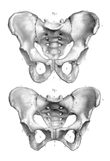
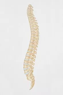
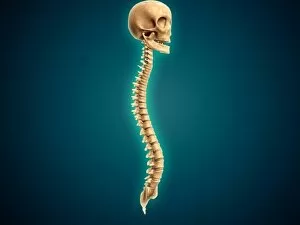
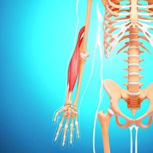
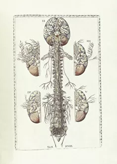
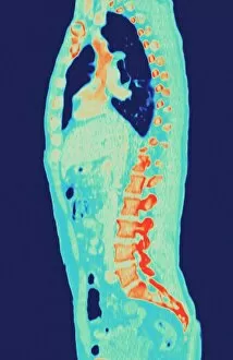
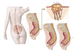
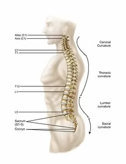
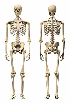
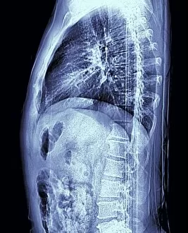
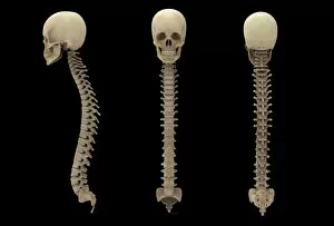
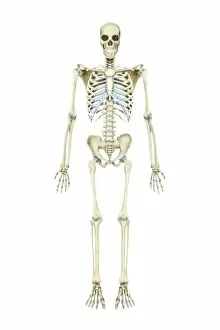
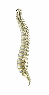
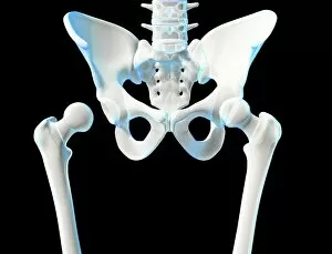
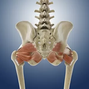
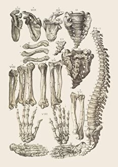
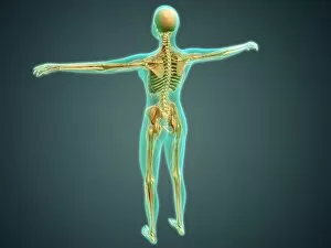
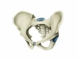
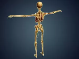
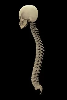
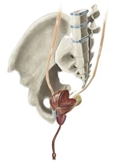
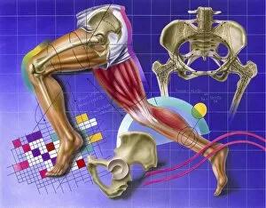
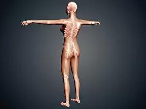
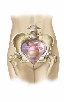
The coccyx, also known as the tailbone, is a small triangular bone located at the base of the spine. It may seem insignificant in size, but it plays a crucial role in providing support and stability to our body. Dating back to 1896, an engraving of male and female pelvis showcases the intricate structure surrounding the coccyx. This illustration highlights how this bone fits snugly between the hip bones, forming part of our pelvic region. A side view diagram from 1866 reveals the human spine's complexity, with each vertebra carefully aligned. Among them lies our coccyx – a vital component that contributes to maintaining proper posture and balance. Intriguing conceptual imagery depicts a human skull alongside its spinal cord. This representation emphasizes how interconnected these two structures are; even though they differ greatly in appearance, they share an intimate relationship within our bodies. Artwork from 1810 beautifully captures the musculature of a human arm. While seemingly unrelated to the coccyx at first glance, it reminds us that every part of our body is intricately connected through muscles and bones working together harmoniously. Bartholomeo Eustachi's masterpiece "The Science of Human Anatomy" delves into further detail about various anatomical features - including an exploration of the coccyx - shedding light on its significance within our overall skeletal framework. Modern medical advancements have allowed for detailed MRI scans showcasing normal torsos. These images provide valuable insights into not only understanding healthy anatomy but also highlighting any potential abnormalities or injuries affecting structures like the coccyx. An anterior view displays both normal and arthritic pelvises with hip bones clearly visible. The former demonstrates what we aspire towards – optimal health and function – while contrasting against arthritis-induced changes that can impact mobility and quality of life for individuals affected by such conditions. Lastly, an anterior view image labels different components comprising our skeletal system.