
Endoscopic Collection

"Exploring the Hidden World: Endoscopic Insights into Ovarian Cysts, Uterus

Background imageEndoscopic Collection: Ovarian cyst, endoscope view C017 / 6800

Ovarian cyst, endoscope view C017 / 6800
Ovarian cyst. Endoscope (laparascope) view of a cyst (white) on a patients ovary. Ovarian cysts are common in women of child-bearing age

Background imageEndoscopic Collection: Uterus, endoscope view C017 / 6805

Uterus, endoscope view C017 / 6805
Uterus. Endoscope (laparascope) view of a healthy uterus (womb, large, upper centre), with the two fallopian tubes (left and right) and an ovary (white, lower right)

Background imageEndoscopic Collection: Gastric antral vascular ectasia C016 / 8328

Gastric antral vascular ectasia C016 / 8328
Gastric antral vascular ectasia. Endoscopic view of the antrum (final portion of stomach), affected by gastric antral vascular ectasia (GAVE)

Background imageEndoscopic Collection: Colon cancer, endoscopic view C016 / 8308

Colon cancer, endoscopic view C016 / 8308
Colon cancer. Endoscopic view of a malignant (cancerous) adenocarcinoma (upper right) in a patients ascending colon (large intestine)

Background imageEndoscopic Collection: Severe gastritis, endoscope view C016 / 3898

Severe gastritis, endoscope view C016 / 3898
Severe gastritis, endoscope view. Gastritis is inflammation of the stomach lining. It causes pain on eating or drinking, and may cause nausea and vomiting

Background imageEndoscopic Collection: Ovarian cyst, endoscope view C017 / 6801

Ovarian cyst, endoscope view C017 / 6801
Ovarian cyst. Endoscope (laparascope) view of a cyst (white) on a patients ovary. Ovarian cysts are common in women of child-bearing age

Background imageEndoscopic Collection: Ulcerative pancolitis C018 / 0928

Ulcerative pancolitis C018 / 0928
Ulcerative pancolitis. Endoscope view of the appendix aperture in the caecum (start of the large intestine) of an 11 year old patient with ulcerative pancolitis showing inflamed mucosa

Background imageEndoscopic Collection: Ovary, endoscope view C017 / 6803

Ovary, endoscope view C017 / 6803
Ovary. Endoscope (laparascope) view of a healthy ovary (white). There are two ovaries in the female body, which release an egg (ovum) during the monthly ovulation cycle

Background imageEndoscopic Collection: Ovarian cyst, endoscope view C017 / 6802

Ovarian cyst, endoscope view C017 / 6802
Ovarian cyst. Endoscope (laparascope) view of a cyst (white) on a patients ovary. Ovarian cysts are common in women of child-bearing age

Background imageEndoscopic Collection: Ectopic pregnancy, endoscope view C017 / 6804

Ectopic pregnancy, endoscope view C017 / 6804
Ectopic pregnancy. Endoscope (laparascope) view of an ectopic pregnancy in a patients fallopian tube. An ectopic pregnancy (eccysis)

Background imageEndoscopic Collection: Pouchitis, endoscope view C015 / 5074

Pouchitis, endoscope view C015 / 5074
Pouchitis, endoscope view. This is the inflammation of an artificial rectum, known as an ileal pouch, formed in patients that have had their large intestine removed

Background imageEndoscopic Collection: Thorascopic surgery, artwork C016 / 6842

Thorascopic surgery, artwork C016 / 6842
Thorascopic surgery. Artwork of an overhead view of surgeons carrying out video-assisted thoracoscopic surgery (VATS). This is a form of keyhole (minimally invasive) endoscopic surgery

Background imageEndoscopic Collection: Colon cancer, barium contrast CT scan

Colon cancer, barium contrast CT scan
Colon cancer. Coloured barium contrast computed tomography (CT) scan, and 3D CT colonoscopy image (inset), of an axial section through the sigmoid colon (large intestine) of a 54-year-old patient

Background imageEndoscopic Collection: Colon cancer, 3D colonoscopy image

Colon cancer, 3D colonoscopy image
Colon cancer. 3D colonoscopy image showing the location of a cancerous growth (centre right) in the colon of a 59-year-old patient. Here, the growth has caused a narrowing (stenosis) of the colon

Background imageEndoscopic Collection: Colon polyp, 3D colonoscopy image

Colon polyp, 3D colonoscopy image
Colon polyp. Coloured 3D computed tomography (CT) colonoscopy image of a pedunculated (stalked) polyp in the sigmoid colon (large intestine) of a 50-year-old patient

Background imageEndoscopic Collection: Colon cancer, CT scans

Colon cancer, CT scans
Colon cancer. 2D computed tomography (CT) rendering (left) and 3D colonoscopy image (right) showing the location of a cancerous growth in the colon of a 59-year-old patient

Background imageEndoscopic Collection: Bowel disease in the colon C016 / 8338

Bowel disease in the colon C016 / 8338
Bowel disease in the colon. Endoscopic view of diverticular disease (diverticulosis) affecting the sigmoid colon. Diverticulosis causes sacs (diverticula)

Background imageEndoscopic Collection: Secondary bowel cancer, endoscopic view C016 / 8337

Secondary bowel cancer, endoscopic view C016 / 8337
Secondary bowel cancer. Endoscopic view of the interior of a patients descending duodenum (first part of the small intestine), showing cancer that has spread (metastasised) from the pancreas

Background imageEndoscopic Collection: Intestinal lymphoid hyperplasia C016 / 8334

Intestinal lymphoid hyperplasia C016 / 8334
Intestinal lymphoid hyperplasia. Endoscopic view of lymphoid hyperplasia in the terminal ileum - the last part of the small intestine

Background imageEndoscopic Collection: Hiatal hernia, endoscopic view C016 / 8330

Hiatal hernia, endoscopic view C016 / 8330
Hiatal hernia. Endoscopic view of a hiatal hernia (gastro-oesophageal prolapse), seen from below, with the endoscope(black) seen in an inverted position inside the stomach

Background imageEndoscopic Collection: Hiatal hernia, endoscopic view C016 / 8329

Hiatal hernia, endoscopic view C016 / 8329
Hiatal hernia. Endoscopic view of a hiatal hernia (gastro-oesophageal prolapse), seen from above. A hernia is the protrusion of an organ, or the fascia of an organ

Background imageEndoscopic Collection: Hyperplastic polyps in the stomach C016 / 8327

Hyperplastic polyps in the stomach C016 / 8327
Hyperplastic polyps in the stomach. Endoscopic view of small hyperplastic polyps (growths) in the gastric fundus of the stomach

Background imageEndoscopic Collection: Major duodenal papilla, endoscopic view C016 / 8324

Major duodenal papilla, endoscopic view C016 / 8324
Major duodenal papilla. Endoscopic view of the interior of a persons small intestine, showing the major duodenal papilla. This structure is where the common bile duct

Background imageEndoscopic Collection: Small intestine, endoscopic view C016 / 8323

Small intestine, endoscopic view C016 / 8323
Small intestine. Endoscopic view of the healthy interior of a persons duodenum - the first part of the small intestine, showing the muscular wall

Background imageEndoscopic Collection: Lining of the duodenum, endoscopic view C016 / 8321

Lining of the duodenum, endoscopic view C016 / 8321
Lining of the duodenum. Endoscopic view of the lining of the duodenum - the first section of the small intestine - showing lipid (fat, white) filled villi

Background imageEndoscopic Collection: Intestinal polyp removal, endoscopic view C016 / 8325

Intestinal polyp removal, endoscopic view C016 / 8325
Intestinal polyp removal. Endoscopic view of a polyp (centre-right) being removed from the sigmoid colon (large intestine). Polyps are benign (non-cancerous) growths that arise from mucous membranes

Background imageEndoscopic Collection: Diverticulum of the duodenum C016 / 8320

Diverticulum of the duodenum C016 / 8320
Diverticulum of the duodenum. Endoscope view of a diverticulum in the wall of the descending part of the duodenum - the first section of the small intestine

Background imageEndoscopic Collection: Angiodysplasia, endoscopic view C016 / 8318

Angiodysplasia, endoscopic view C016 / 8318
Angiodysplasia. Endoscopic view of the interior of a patients caecum (beginning of the large intestine), showing a small angiodysplasia

Background imageEndoscopic Collection: Crohns disease, endoscopic view

Crohns disease, endoscopic view
Crohns disease. Endoscopic view of the interior of a patients neoterminal ileum affected by Crohns disease, showing ulceration (white)

Background imageEndoscopic Collection: Ileocaecal valve, endoscopic view C016 / 8317

Ileocaecal valve, endoscopic view C016 / 8317
Ileocaecal valve. Endoscopic view of the healthy interior of a persons ileum (small intestine), showing the ileocaecal valve

Background imageEndoscopic Collection: Ulcerative colitis, endoscopic view C016 / 8313

Ulcerative colitis, endoscopic view C016 / 8313
Ulcerative colitis. Endoscopic view of the interior of a patients distal colon (large intestine) affected by ulcerative colitis (UC), showing ulceration (white areas)

Background imageEndoscopic Collection: Antrum occult mucosal bleeding C016 / 8314

Antrum occult mucosal bleeding C016 / 8314
Antrum occult mucosal bleeding. Endoscopic view of the interior of a patients antrum (final portion of stomach), showing bleeding (red) of the mucosal lining

Background imageEndoscopic Collection: Ulcerative colitis, endoscopic view C016 / 8312

Ulcerative colitis, endoscopic view C016 / 8312
Ulcerative colitis. Endoscopic view of the interior of a patients distal colon (large intestine) affected by ulcerative colitis (UC), showing ulceration (white areas)

Background imageEndoscopic Collection: Ulcerative proctitis, endoscopic view C016 / 8311

Ulcerative proctitis, endoscopic view C016 / 8311
Ulcerative colitis. Endoscopic view of the interior of a patients anus, showing ulceration (white areas) cause by ulcerative proctitis

Background imageEndoscopic Collection: Ulcerative colitis, endoscopic view C016 / 8310

Ulcerative colitis, endoscopic view C016 / 8310
Ulcerative colitis. Endoscopic view of the interior of a patients colon (large intestine) affected by ulcerative colitis (UC), showing ulceration (white areas)

Background imageEndoscopic Collection: Ulcerative colitis, endoscopic view C016 / 8309

Ulcerative colitis, endoscopic view C016 / 8309
Ulcerative colitis. Endoscopic view of the interior of a patients colon (large intestine) affected by ulcerative colitis (UC), showing ulceration (white areas)

Background imageEndoscopic Collection: Ulcerative colitis, endoscopic view C016 / 8306

Ulcerative colitis, endoscopic view C016 / 8306
Ulcerative colitis. Endoscopic view of the interior of a patients distal colon (large intestine) affected by ulcerative colitis (UC), showing ulceration (white areas)

Background imageEndoscopic Collection: Colon cancer, endoscopic view C016 / 8307

Colon cancer, endoscopic view C016 / 8307
Colon cancer. Endoscopic view of a malignant (cancerous) adenocarcinoma (lower right) in a patients ascending colon (large intestine)

Background imageEndoscopic Collection: Melanosis coli, endoscopic view C016 / 8304

Melanosis coli, endoscopic view C016 / 8304
Melanosis coli. Endoscopic view of the interior of a patients colon affected by melanosis coli (pseudomelanosis coli). This benign condition causes brown pigmentation of the lining of the colon

Background imageEndoscopic Collection: Capsule endoscopy

Capsule endoscopy. Doctor holding a capsule endoscope. This pill-shaped capsule contains a small camera and is swallowed by a patient to provide real-time video images of the stomach and intestines


For sale as Licensed Images

Choose your image, Select your licence and Download the media

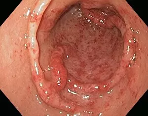
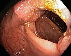
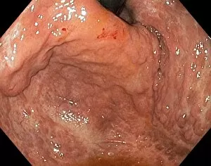
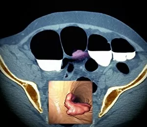
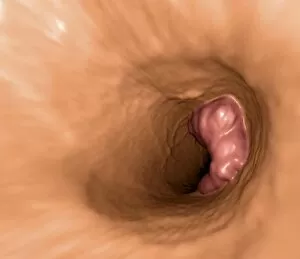
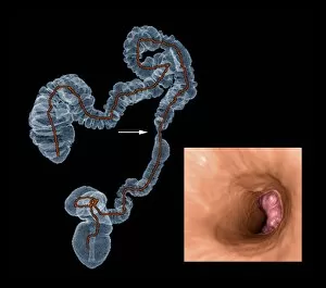
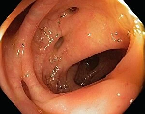
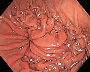
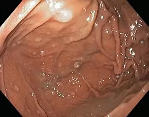
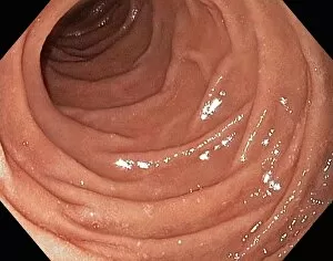
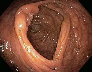
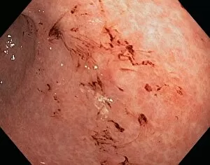
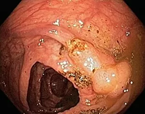
"Exploring the Hidden World: Endoscopic Insights into Ovarian Cysts, Uterus, and Gastric Antral Vascular Ectasia" Step into the fascinating realm of endoscopy as we delve deep into the intricate details of various medical conditions. Witness the mesmerizing views captured through an endoscope lens, revealing a world unseen by the naked eye. Firstly, behold the enigmatic ovarian cysts as they appear under endoscope view C017/6800. These fluid-filled sacs within the ovaries can cause discomfort and require careful examination for proper diagnosis and treatment. Moving on to explore further, let us observe another captivating glimpse of a uterus under endoscope view C017/6805. This remarkable tool allows physicians to closely examine this vital reproductive organ, aiding in detecting abnormalities or potential issues that may affect fertility or overall health. Venturing deeper down our exploration path is an intriguing encounter with gastric antral vascular ectasia (GAVE) showcased in image C016/8328. This condition presents itself with dilated blood vessels in the stomach lining, often causing gastrointestinal bleeding and necessitating precise intervention using endoscopic techniques. Continuing our journey through this hidden landscape are more instances of ovarian cysts depicted in images C017/6801 and C017/6802. These visualizations provide invaluable insights for medical professionals striving to understand these common yet complex growths that can impact women's health. Next up is a rare but critical condition known as ectopic pregnancy displayed via endoscope view C017/6804. The ability to visualize such cases aids doctors in diagnosing this potentially life-threatening situation where fertilized eggs implant outside of the uterus. Shifting gears slightly towards inflammatory bowel diseases brings us face-to-face with pouchitis portrayed through endoscope view C015/5074. By examining this inflammation occurring after ileal pouch-anal anastomosis surgery, endoscopy plays a pivotal role in managing this challenging condition.