
Human Ear Collection
The human ear is an intricate and fascinating organ that plays a vital role in our ability to hear and perceive sound


For sale as Licensed Images
Choose your image, Select your licence and Download the media
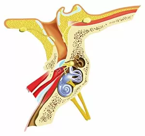
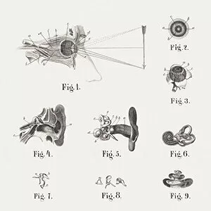
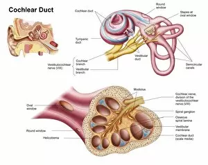
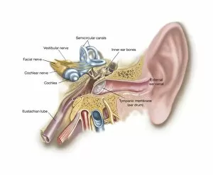
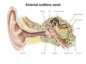
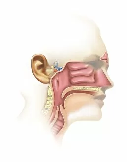
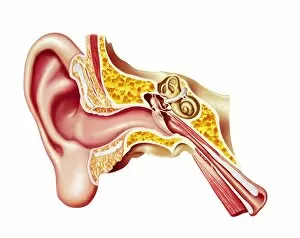
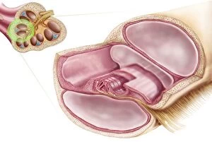
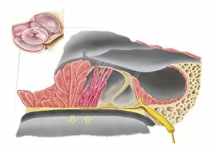
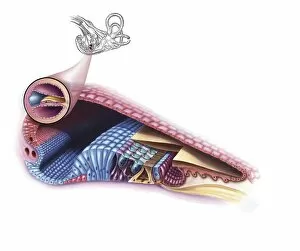
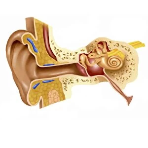
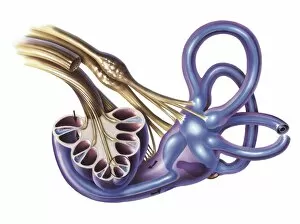
The human ear is an intricate and fascinating organ that plays a vital role in our ability to hear and perceive sound. A cross-section diagram of the human ear reveals its complex structure, consisting of various parts such as the auditory canal, eardrum, semicircular canals, cochlea, cochlear nerve, and eustachian tube. Scientific illustrations depicting the anatomy of the ear provide us with a visual understanding of this remarkable sensory system. These illustrations showcase the inner workings of the hearing organ and highlight its importance among our senses. One captivating illustration from 1861 showcases both the eye and ear's anatomical details side by side. This historical artwork reminds us of how far we have come in understanding these intricate structures over time. Another illustration focuses specifically on the cochlear duct within the human ear. The delicate intricacies displayed in this image emphasize just how finely tuned our hearing capabilities are. Moving towards external features, an illustration highlights the external auditory canal labeled for better comprehension. This depiction allows us to visualize how sound waves enter our ears before reaching deeper into their internal mechanisms. In a more lighthearted portrayal, a cartoon featuring a boy holding a large conch shell against his ear adds humor to our exploration of this topic. With fish swimming out from one end while water flows from another, it playfully demonstrates how sounds can be distorted or amplified depending on their environment. Expanding beyond humans' realm but still relevant to auditory systems is an intriguing cross-section illustration showcasing an Earwig insect inside an auditory canal touching its tympanic membrane with antennae. This unique perspective sheds light on different organisms' adaptations for hearing purposes. Lastly, exploring not only hearing but also sinuses connected to it provides further insight into related anatomical structures that contribute to overall balance and equilibrium perception. These captivating images and diagrams offer glimpses into both scientific knowledge about human ears throughout history as well as imaginative interpretations of their functions.