
Lungs Collection (#2)
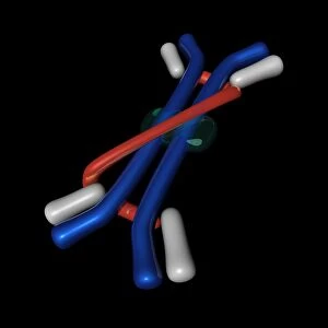
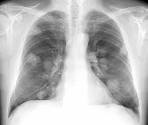
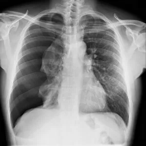
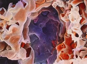
"Lungs: The Vital Organ Responsible for Life-Sustaining Breath" Front View of Lungs: Behold the intricate beauty of our lungs


For sale as Licensed Images
Choose your image, Select your licence and Download the media
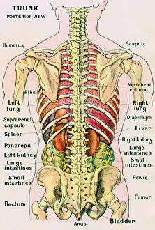
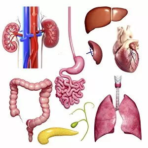
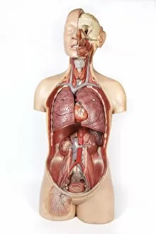
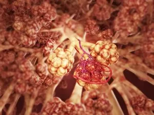
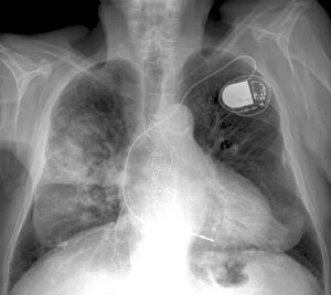
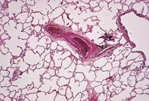
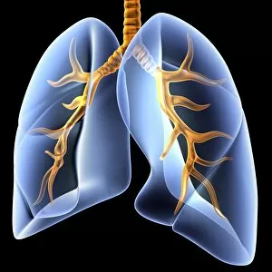
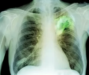
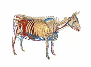
"Lungs: The Vital Organ Responsible for Life-Sustaining Breath" Front View of Lungs: Behold the intricate beauty of our lungs, a masterpiece of nature that fuels every breath we take. Diagram of the Lungs and Bronchial Tubes: Explore the complex network within our respiratory system, where oxygen is exchanged for carbon dioxide in a remarkable process. Tuberculosis X-ray: Unveiling the silent threat - tuberculosis, an infectious disease that can affect these delicate organs and disrupt their function. Diagram of the Heart, Lungs, and Windpipe: Witness how these vital components work together seamlessly to ensure proper oxygenation throughout our body. Pig Anatomy Artwork: Discover surprising similarities between human and pig lungs as we delve into comparative anatomy through captivating artwork. Diagram of Human Lungs Showing Blood Supply: Marvel at the intricate web of blood vessels nourishing our lungs with life-giving nutrients while removing waste products. Tension Pneumothorax X-ray: A glimpse into a medical emergency - tension pneumothorax, where air accumulates in the chest cavity causing lung collapse and compromising breathing. Heart and Lungs: A Synchronized Symphony Witness how these two powerhouses collaborate harmoniously to pump oxygen-rich blood throughout our body's vast network. Diagram of the Lungs (Front View): Delve deeper into understanding this essential organ from a frontal perspective, appreciating its complexity like never before. Cystic Fibrosis: Battling Against Lung Disease Learn about cystic fibrosis—a genetic disorder affecting lung function—and explore ongoing research aiming to improve patients' quality of life. Konstantin Buteyko: Soviet Doctor Revolutionizing Breathing Techniques Discover how Dr Konstantin Buteyko's innovative methods have helped countless individuals optimize their lung capacity through specialized breathing exercises. Human Venous System.