
Organ Of Corti Collection
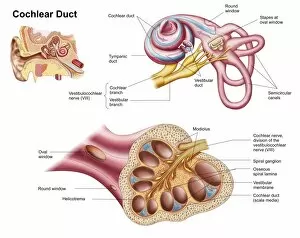
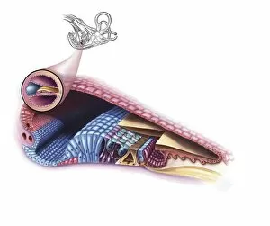
The Organ of Corti


For sale as Licensed Images
Choose your image, Select your licence and Download the media
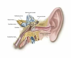
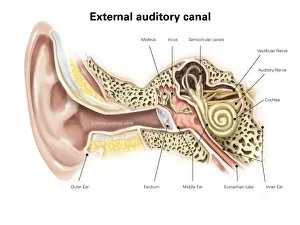
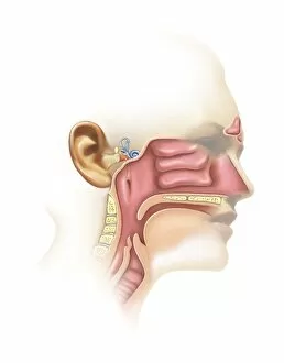
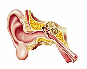
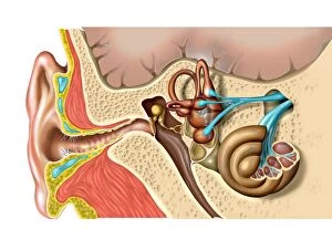
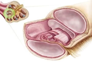
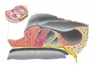
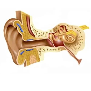
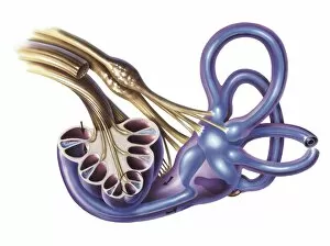
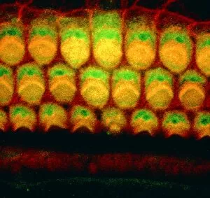
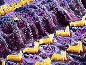
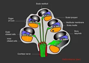
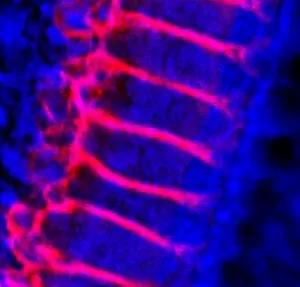
The Organ of Corti: Unveiling the Intricacies of the Inner Ear Step into the fascinating world of human anatomy with scientific illustrations that unravel the mysteries of our auditory system. Explore the intricate structures that make up this remarkable sensory organ, known as the Organ of Corti. Delving deep into the cochlear duct, we discover a complex network within our ear. The external auditory canal, labeled meticulously in these illustrations, serves as a gateway to this extraordinary realm. As we venture further, we encounter inner ear hairs captured in stunning detail through scanning electron microscopy (SEM). These delicate hairs play a vital role in converting sound vibrations into electrical signals that our brain can interpret. Examining both the anatomy and sinuses within this region allows us to appreciate its complexity fully. Cutaway diagrams provide an insightful glimpse into how all these components fit together seamlessly to create our sense of hearing. At last, we arrive at our main protagonist – the Organ of Corti itself. Digital illustrations vividly portray this essential structure found within the cochlea. Acting as nature's own orchestra conductor, it orchestrates an exquisite symphony by transforming sound waves into nerve impulses for transmission to our brain. Medical illustrations reveal another crucial element - endolymph in the membranous labyrinth. This fluid-filled space enhances sound conduction and ensures optimal functioning of this miraculous organ. As we explore every nook and cranny inside this interior wonderland called Cochlea, one thing becomes abundantly clear – each component has a purpose; each structure contributes to our ability to hear and perceive sound accurately. So let us marvel at these captivating scientific illustrations that allow us to peer beneath layers unseen by naked eyes. Let us celebrate humanity's quest for knowledge as we uncover more about ourselves through understanding organs like Corti - unlocking secrets hidden deep within their anatomical intricacies.