Pelvis Collection (#2)

The pelvis: A vital foundation of the human body's intricate design

Background imagePelvis Collection: Midwifery, illustration from Encyclopedia Britannica, published in Edinburgh

Midwifery, illustration from Encyclopedia Britannica, published in Edinburgh
DGC398035 Midwifery, illustration from Encyclopedia Britannica, published in Edinburgh, 1798 (copperplate engraving) by Bell, Alexander (fl)

Background imagePelvis Collection: Two studies of a hanging skeleton (pen & ink with wash)

Two studies of a hanging skeleton (pen & ink with wash)
AII80166 Two studies of a hanging skeleton (pen & ink with wash) by Vinci, Leonardo da (1452-1519); Gabinetto dei Disegni e Stampe, Galleria Degli Uffizi, Florence, Tuscany, Italy; Italian

Background imagePelvis Collection: Normal posterior view of the hip bones and gluteus maximus muscle including the lumbar

Normal posterior view of the hip bones and gluteus maximus muscle including the lumbar
Detail, Anatomy, Diagram, Muscle, Illustration, Bone, Medical, Hip, Biology, Pelvis, Femur, See Through, Rear View, Cut Out, White Background, Artwork, Color Image, Close Up, Healthcare And Medicine

Background imagePelvis Collection: Normal posterior view of the back highlighting the multifidus muscle

Normal posterior view of the back highlighting the multifidus muscle
Detail, Anatomy, Diagram, Muscle, Illustration, Ribs, Bone, Medical, Biology, Pelvis, See Through, Rear View, Cut Out, White Background, Artwork, Color Image, Close Up, Healthcare And Medicine

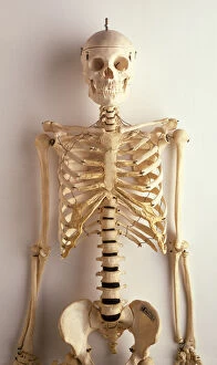
Background imagePelvis Collection: Human rib cage, jaw bones, neck vertabrae leading to sternum, collar bones and ribcage

Human rib cage, jaw bones, neck vertabrae leading to sternum, collar bones and ribcage, spinal column continuing to pelvis, front view

Background imagePelvis Collection: Anatomy skeleton

Anatomy skeleton
Diagram of the human skeleton from front and back

Background imagePelvis Collection: Female body with bone skeleton and internal organs superimposed

Female body with bone skeleton and internal organs superimposed
Anatomy of female body with bone skeleton and all internal organs superimposed

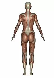
Background imagePelvis Collection: Anatomy of female muscular system, back view

Anatomy of female muscular system, back view

Background imagePelvis Collection: Anterior view of human pelvis, with labels

Anterior view of human pelvis, with labels

Background imagePelvis Collection: Visualization of human diaphragm

Visualization of human diaphragm

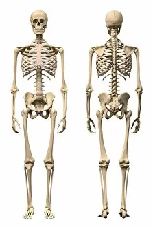
Background imagePelvis Collection: Anatomy of male human skeleton, front view and back view

Anatomy of male human skeleton, front view and back view

Background imagePelvis Collection: Human midsection with internal organs

Human midsection with internal organs

Background imagePelvis Collection: Human musculature, artwork F007 / 2038

Human musculature, artwork F007 / 2038
Human musculature, computer artwork

Background imagePelvis Collection: Human arm musculature, artwork F007 / 1379

Human arm musculature, artwork F007 / 1379
Human arm musculature, computer artwork

Background imagePelvis Collection: Dorsogluteal injection site, artwork

Dorsogluteal injection site, artwork
Intramuscular dorsogluteal injection site. Artwork showing the correct site (red square) for a dorsogluteal injection. The dorsogluteal muscle is in the upper outside quadrant of the buttock

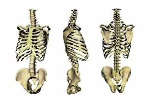
Background imagePelvis Collection: Human skeleton anatomy, artwork

Human skeleton anatomy, artwork
Human skeleton anatomy, computer artwork. Three views of the bones of the human torso, seen from an oblique frontal view (left), from the side (centre) and from the front (right)

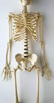
Background imagePelvis Collection: Lower half of human skeleton, jaw, spinal column, rib cage, pelvis, femurs, arms

Lower half of human skeleton, jaw, spinal column, rib cage, pelvis, femurs, arms, hands and fingers, front view

Background imagePelvis Collection: Upper part of human skeleton, skull, spinal column, ribcage, shoulders, collar bones

Upper part of human skeleton, skull, spinal column, ribcage, shoulders, collar bones, upper arms and pelvis, front view

Background imagePelvis Collection: 15th century Costume

15th century Costume
1. Jean de Linden (1394) 2. Duc de Baviere (1430-1470) (whose name is unknown) 3. Albert III, Achilles de Brandebourg (1475) wearing an Electors cap 4. Ludwig von Hutten (1414) 5

Background imagePelvis Collection: The human skeleton engraving 1895

The human skeleton engraving 1895
The human skeleton engraving

Background imagePelvis Collection: Human thorax, shoulder girdle, and pelvis posterior view

Human thorax, shoulder girdle, and pelvis posterior view

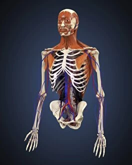
Background imagePelvis Collection: Human upper body showing bones, muscles and circulatory system

Human upper body showing bones, muscles and circulatory system

Background imagePelvis Collection: Rear view of human skeleton showing kidney and nervous system

Rear view of human skeleton showing kidney and nervous system

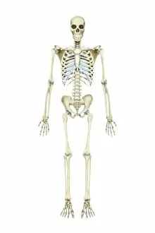
Background imagePelvis Collection: Anterior view of human skeletal system

Anterior view of human skeletal system

Background imagePelvis Collection: Anatomy of female body with skeleton, stylized look

Anatomy of female body with skeleton, stylized look

Background imagePelvis Collection: Abdominal quadrants with internal organs and rib cage

Abdominal quadrants with internal organs and rib cage
Abdominal quadrants, posterior view with internal organs and rib cage

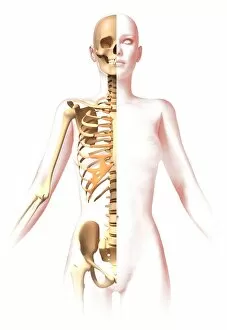
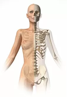
Background imagePelvis Collection: Female body with bone skeleton superimposed

Female body with bone skeleton superimposed
Medical illustration of female body with bone skeleton superimposed

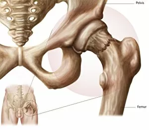
Background imagePelvis Collection: Anatomy of hip fracture

Anatomy of hip fracture

Background imagePelvis Collection: Anatomy of male urinary system

Anatomy of male urinary system

Background imagePelvis Collection: Anatomy of human pelvic bone

Anatomy of human pelvic bone

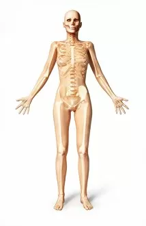
Background imagePelvis Collection: Female standing, with skeletal bones superimposed, front view

Female standing, with skeletal bones superimposed, front view
3D rendering of a naked female standing, with skeletal bones superimposed, front view

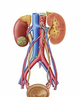
Background imagePelvis Collection: Early stage of kidney cancer with a tumor visible on the kidney

Early stage of kidney cancer with a tumor visible on the kidney

Background imagePelvis Collection: The human skeleton and muscular system, front view

The human skeleton and muscular system, front view
Medical illustration of the human skeleton and muscular system, front view

Background imagePelvis Collection: Anatomy of male body with half skeleton and half muscular system

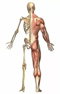
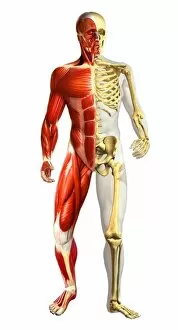
Anatomy of male body with half skeleton and half muscular system
Anatomy of male body with half skeleton and half muscular representations

Background imagePelvis Collection: Human musculature, artwork F007 / 4130

Human musculature, artwork F007 / 4130
Human musculature, computer artwork

Background imagePelvis Collection: Human arm musculature, artwork F007 / 4724

Human arm musculature, artwork F007 / 4724
Human arm musculature, computer artwork

Background imagePelvis Collection: Human musculature, artwork F007 / 4829

Human musculature, artwork F007 / 4829
Human musculature, computer artwork

Background imagePelvis Collection: Pelvic-femoral muscles, 1831 artwork

Pelvic-femoral muscles, 1831 artwork
Pelvic-femoral muscles, superficial layer. Part of the spine at top left. This anatomical artwork is plate 124 from volume 2 (1831) of Traite complet de l anatomie de l homme (1831-1854)

Background imagePelvis Collection: Human musculature, artwork F007 / 3311

Human musculature, artwork F007 / 3311
Human musculature, computer artwork

Background imagePelvis Collection: Hip joint bones and anatomy, artwork C014 / 2032

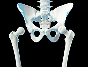
Hip joint bones and anatomy, artwork C014 / 2032
Hip joint bones and anatomy. Artwork of the bones that form the two hip joints (right and left). The hip is a ball-and-socket joint that provides the mobility needed for walking and running

Background imagePelvis Collection: Uterine fibroids, MRI scan C018 / 0466

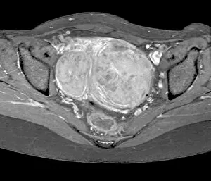
Uterine fibroids, MRI scan C018 / 0466
Uterine fibroids. Magnetic resonance imaging (MRI) scan of the pelvis of a 47 year old female with two large fibroids (centre) in her uterus

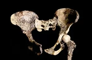
Background imagePelvis Collection: Australopithecus africanus pelvis, STS-14 C015 / 6919

Australopithecus africanus pelvis, STS-14 C015 / 6919
Australopithecus africanus pelvis (STS-14). This fossil specimen was discovered in 1947, in Sterkfontein, South Africa. The entire specimen consists of the pelvis, part of the vertebral column

Background imagePelvis Collection: Coccyx pain, artwork

Coccyx pain, artwork
Coccyx (tailbone) pain, computer artwork

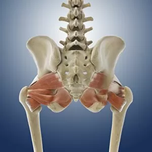
Background imagePelvis Collection: Buttock muscles, artwork C013 / 4414

Buttock muscles, artwork C013 / 4414
Buttock muscles. Computer artwork of a posterior view of the hips showing some of the muscles (red) of the buttocks. These are shown attached to the femurs (thigh bones) and the pelvis bones
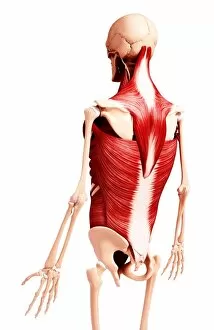
Background imagePelvis Collection: Male torso skeleton

Male torso skeleton. The bones of the chest and abdomen surround and protect the organs and provide support for the body. The ribcage (upper frame) encloses the heart and lungs
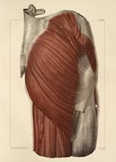
Background imagePelvis Collection: Torso bones

Torso bones. Computer artwork of the bones of the human torso. The backbone (running vertically down the centre) is a flexible column of 26 bones (vertebrae)

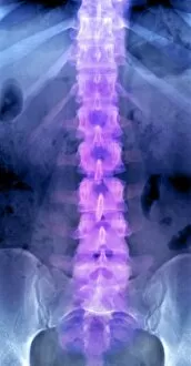
Background imagePelvis Collection: Normal lumbar spine, X-ray

Normal lumbar spine, X-ray
Normal lumbar spine. Coloured frontal X-ray showing bones of the lower (lumbar) spine (centre, purple). The bones of the spine (block-shaped) are called vertebrae

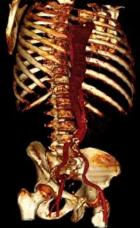
Background imagePelvis Collection: Aortic dissection, 3D CT scan

Aortic dissection, 3D CT scan
Aortic dissection. 3D computed tomography (CT) scan of a 67-year-old man with an aortic dissection. The aorta, the bodys main artery, is running vertically down centre (red)


For sale as Licensed Images

Choose your image, Select your licence and Download the media

The pelvis: A vital foundation of the human body's intricate design. From its role in supporting the backbone, including ribs and pelvis, to its distinct differences between male and female anatomy depicted in a captivating 1896 engraving. Explore the connection between the cardiovascular system and this historical artwork, showcasing the significance of every bone and joint. Witness the transformative power of hip replacement through stunning artistic representations and X-rays capturing both total hip replacements and normal spines. Delve into computer artwork depicting an upper body skeleton, highlighting how each element contributes to our overall structure. Study a detailed diagram showcasing the bones of the right leg and hip, unraveling their complexity with precision. Marvel at Julien Bougle's masterpiece - a human body adorned with superimposed colored plates that reveal hidden wonders within us all. Discover how osteoarthritis affects this crucial region through an enlightening X-ray image. Finally, admire F007/1810's exquisite artwork unveiling the intricacies of human arm musculature – a testament to our remarkable physical capabilities.