
Pulmonology Collection
Pulmonology, the fascinating study of the respiratory system, delves deep into the intricate anatomy and functions of our lungs


For sale as Licensed Images
Choose your image, Select your licence and Download the media
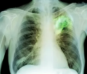
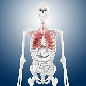
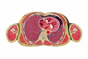
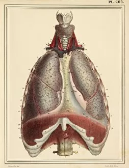
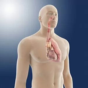
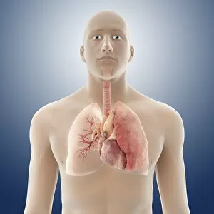
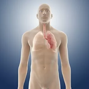
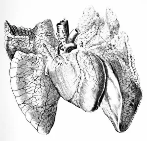
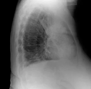
Pulmonology, the fascinating study of the respiratory system, delves deep into the intricate anatomy and functions of our lungs. From stomach anatomy to bronchial tree illustrations, artwork has played a crucial role in unraveling the mysteries of this vital organ. In an 1825 masterpiece, we witness a detailed depiction of heart-lung anatomy, showcasing their interconnectedness and symbiotic relationship. The artist's skillful strokes bring to life the complex network that allows oxygen-rich blood to flow through our bodies. Another captivating artwork showcases a cross-section view of pneumothorax - a condition where air accumulates between the lung and chest wall. This visual representation helps medical professionals understand its impact on lung function and devise effective treatment strategies. The bronchial tree takes center stage in yet another stunning illustration. Its branches spread like delicate tendrils throughout our lungs, ensuring efficient airflow and gas exchange. These artistic renderings help us appreciate the intricacy of this essential respiratory pathway. Artwork featuring lungs captures their beauty while highlighting their critical role in sustaining life. Whether it's vibrant colors or meticulous detailing, these visuals remind us how every breath fuels our existence. An 1825 artwork dedicated solely to bronchial lung anatomy provides valuable insights into this specialized structure within our respiratory system. It unravels its complexities with precision and artistry, aiding pulmonologists in diagnosing conditions affecting these specific regions. Lungs are not solitary entities but work harmoniously with other organs such as the diaphragm - showcased elegantly in an 1825 piece. This collaboration ensures proper breathing mechanics for optimal oxygenation throughout our body systems. Human respiratory system artwork serves as an educational tool for both students and experts alike by presenting a comprehensive overview of its various components working together seamlessly. Such visuals aid understanding from trachea to alveoli – each playing a unique role in respiration.