Histological Collection (#3)

"Exploring the Intricacies of Histological Wonders: Unveiling the Hidden Beauty within Our Bodies" Step into the fascinating world of histology

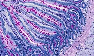
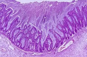
Background imageHistological Collection: Small intestine, light micrograph

Small intestine, light micrograph
Small intestine. Light micrograph of a section through the small intestine. The finger-like projections at left are the villi. These increase the surface area for the absorption of food

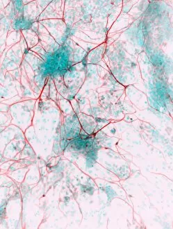
Background imageHistological Collection: Stem cell-derived neurons, micrograph

Stem cell-derived neurons, micrograph
Stem cell-derived neurons. Light micrograph of human nerve cells (neurons) that have been derived from induced pluripotent stem cells (IPS). Tuj1 proteins are cyan, and cell nuclei are red

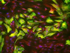
Background imageHistological Collection: Astrocyte brain cells, light micrograph

Astrocyte brain cells, light micrograph
Astrocyte brain cells. Fluorescence light micrograph of primary astrocyte cells from the brain of a mouse. Astrocytes have numerous branches of connective tissue that provide support

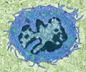
Background imageHistological Collection: Intestinal gland cell, TEM C014 / 1440

Intestinal gland cell, TEM C014 / 1440
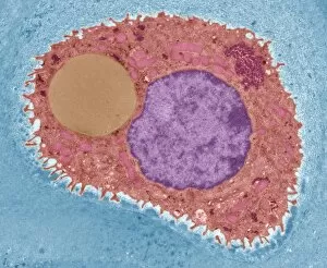
Intestinal gland cell. Transmission electron micrograph (TEM) of a section through an undifferentiated cell from the intestinal tract, showing extensive rough endoplasmic reticulum (ER)

Background imageHistological Collection: Cartilage cell, TEM C014 / 1432

Cartilage cell, TEM C014 / 1432
Cartilage cell. Transmission electron micrograph (TEM) of a section through a chondrocyte cell from hyaline cartilage of the trachea (windpipe)

Background imageHistological Collection: Light microscope C014 / 1407

Light microscope C014 / 1407
Light microscope, seen in silhouetted profile. This microscope has a binocular eyepiece (an eyepiece for each eye). The sample is placed on a glass slide on the mount below centre

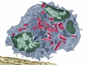
Background imageHistological Collection: Eosinophil white blood cell, TEM C014 / 1437

Eosinophil white blood cell, TEM C014 / 1437
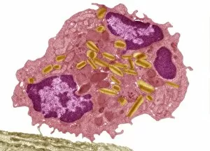
Eosinophil white blood cell. Transmission electron micrograph (TEM) of a section through an eosinophil. Eosinophils, like all white blood cells, are part of the bodys immune system

Background imageHistological Collection: Brain nerve cells, TEM C014 / 0357

Brain nerve cells, TEM C014 / 0357
Brain nerve cells. Transmission electron micrograph (TEM) of a section through brain tissue from the cerebral cortex, showing numerous neurons (nerve cells) surrounded by axons and dendrites

Background imageHistological Collection: Eosinophil white blood cell, TEM C014 / 1439

Eosinophil white blood cell, TEM C014 / 1439
Eosinophil white blood cell. Transmission electron micrograph (TEM) of a section through an eosinophil. Eosinophils, like all white blood cells, are part of the bodys immune system

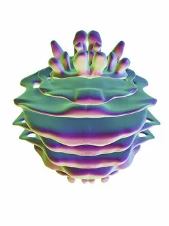
Background imageHistological Collection: Golgi apparatus, artwork F006 / 9211

Golgi apparatus, artwork F006 / 9211
Computer artwork of the Golgi apparatus of the human cell. This organelle functions as a central delivery system for the cell

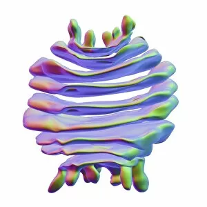
Background imageHistological Collection: Golgi apparatus, artwork F006 / 9199

Golgi apparatus, artwork F006 / 9199
Computer artwork of the Golgi apparatus of the human cell. This organelle functions as a central delivery system for the cell

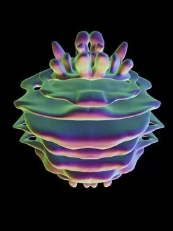
Background imageHistological Collection: Golgi apparatus, artwork F006 / 9189

Golgi apparatus, artwork F006 / 9189
Computer artwork of the Golgi apparatus of the human cell. This organelle functions as a central delivery system for the cell

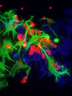
Background imageHistological Collection: Stem cell-derived astrocyte brain cells

Stem cell-derived astrocyte brain cells
Stem cell-derived nerve cells. Fluorescence light micrograph of astrocyte brain cells that have been derived from neural (nerve) stem cells from a mouse

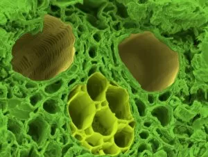
Background imageHistological Collection: Bamboo stem, SEM C015 / 5073

Bamboo stem, SEM C015 / 5073
Bamboo stem. Coloured environmental scanning electron micrograph (ESEM) of a section through the stem of a bamboo plant (family Gramineae), showing a vascular bundle

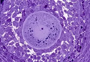
Background imageHistological Collection: Oocyte, light micrograph

Oocyte, light micrograph
Oocyte. Light micrograph of a section through an oocyte within a early antral follicle in an ovary. Oocytes are immature ova, or egg cells

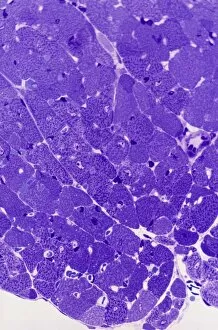
Background imageHistological Collection: Cardiac muscle, light micrograph

Cardiac muscle, light micrograph
Cardiac muscle. Light micrograph of a section through muscle tissue from a heart, showing the cardiomyocyte cells. Heart muscle cells show central nuclei but the dominant feature is the abundance of

Background imageHistological Collection: Colitis, light micrograph

Colitis, light micrograph
Colitis. Light micrograph of a section through colon (large intestine) tissue affected by bacterial infection, causing injury to the mucosal lining, which has resulted in inflammation (colitis)

Background imageHistological Collection: Hairy scalp skin, light micrograph

Hairy scalp skin, light micrograph
Hairy scalp skin. Light micrograph of a section through healthy skin from a human scalp, showing the numerous hair follicles. The scalp is an example of hairy skin

Background imageHistological Collection: Oxalosis, light micrograph

Oxalosis, light micrograph
Oxalosis. Light micrograph of a section through adult bone tissue affected by oxalosis (primary hyperoxaluria), showing mature bone (green)

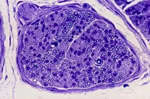
Background imageHistological Collection: Peripheral nerve, light micrograph

Peripheral nerve, light micrograph
Peripheral nerve. Light micrograph of a section through a peripheral nerve. This is a mixed nerve with myelinated axons (dark blue circles)

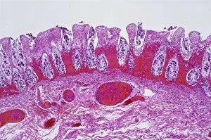
Background imageHistological Collection: Ischaemic bowel, light micrograph

Ischaemic bowel, light micrograph
Ischaemic bowel. Light micrograph of a section through a portion of intestine that has been damaged by an interruption of the blood supply, or ischaemia

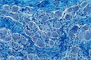
Background imageHistological Collection: Nerve ganglion, light micrograph

Nerve ganglion, light micrograph
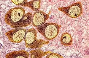
Nerve ganglion. Light micrograph of a section through a dorsal (sensory) spinal root ganglion associated with a sensory nerve root of the spinal cord. Sensory information from peripheral sites e.g

Background imageHistological Collection: Myelinated nerve, light micrograph

Myelinated nerve, light micrograph
Myelinated nerve. Light micrograph of a section through a peripheral myelinated nerve, showing many individual axons each covered with deep blue-staining myelin sheaths

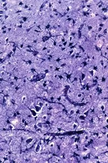
Background imageHistological Collection: Glial cells, light micrograph

Glial cells, light micrograph
Glial cells in the brain. Light micrograph of a section through glial cells (dark) in the grey matter of the brain. Due to their star shape these glial cells are called astrocytes

Background imageHistological Collection: Algae cell wall, SEM

Algae cell wall, SEM
Algae cell wall. Coloured scanning electron micrograph (SEM) of cellulose microfibrils (strands) in a Chaetomorpha sp. algal cell wall

Background imageHistological Collection: Cirrhosis of the liver, light micrograph

Cirrhosis of the liver, light micrograph
Cirrhosis of the liver. Light micrograph of a section through liver tissue affected by cirrhosis, showing a cluster of hepatocyte cells (orange)

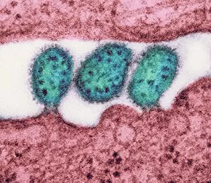
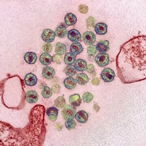
Background imageHistological Collection: Lassa virus particles, TEM C016 / 9409

Lassa virus particles, TEM C016 / 9409
Lassa virus particles. Transmission electron micrograph (TEM) of Lassa virus particles (virions, blue) amongst cell debris. This Arenavirus is the cause of Lassa fever

Background imageHistological Collection: Acute haemorrhagic conjunctivitis, TEM C016 / 9387

Acute haemorrhagic conjunctivitis, TEM C016 / 9387
Acute haemorrhagic conjunctivitis. Transmission electron micrograph (TEM) of a sample of tissue from the conjunctiva of a human eye that is infected by enterovirus particles (purple)

Background imageHistological Collection: Eastern equine encephalitis, TEM C016 / 9390

Eastern equine encephalitis, TEM C016 / 9390
Eastern equine encephalitis. Transmission electron micrograph (TEM) of a section through tissue from a human central nervous system (CNS)

Background imageHistological Collection: Colorado tick fever virus infection, TEM C016 / 9384

Colorado tick fever virus infection, TEM C016 / 9384
Colorado tick fever virus infection. Transmission electron micrograph (TEM) of human tissue infected by Colorado tick fever virus (CTFV) particles (virions, dark circles)

Background imageHistological Collection: Swine flu virus particles, TEM C016 / 9399

Swine flu virus particles, TEM C016 / 9399
Swine flu virus particles. Transmission electron micrograph (TEM) of a section through H3N2 influenza A virus particles (virions)

Background imageHistological Collection: Swine flu virus particles, TEM C016 / 9406

Swine flu virus particles, TEM C016 / 9406
Influenza virus particles. Transmission electron micrograph (TEM) of influenza (flu) virus particles (virions). Each virion consists of ribonucleic acid (RNA, dark patches)

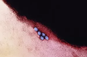
Background imageHistological Collection: HIV virus particles, TEM C016 / 9404

HIV virus particles, TEM C016 / 9404
HIV virus particles. Transmission electron micrograph (TEM) of a section through human immunodeficiency virus (HIV) particles (virions, round)

Background imageHistological Collection: HIV virus particles, TEM C016 / 9405

HIV virus particles, TEM C016 / 9405
HIV virus particles. Transmission electron micrograph (TEM) of a section through human immunodeficiency virus (HIV) particles (virions, round)

Background imageHistological Collection: Swine flu virus particles, TEM C016 / 9400

Swine flu virus particles, TEM C016 / 9400
Swine flu virus particles. Transmission electron micrograph (TEM) of a section through H3N2 influenza A virus particles (virions)

Background imageHistological Collection: Lassa virus particles, TEM C016 / 9408

Lassa virus particles, TEM C016 / 9408
Lassa virus particles. Transmission electron micrograph (TEM) of Lassa virus particles (virions, green) amongst cell debris. This Arenavirus is the cause of Lassa fever

Background imageHistological Collection: Hartmannella vermiformis protozoa cysts C016 / 9402

Hartmannella vermiformis protozoa cysts C016 / 9402
Hartmannella vermiformis protozoa cysts. Transmission electron micrograph (TEM) of a section through cysts (round) containing Hartmannella vermiformis protozoa. H

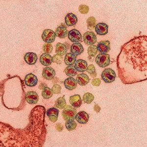
Background imageHistological Collection: Ganjam virus infection, TEM C016 / 9396

Ganjam virus infection, TEM C016 / 9396
Ganjam virus infection. Transmission electron micrograph (TEM) of a section through human tissue infected with ganjam virus particles (virions, blue and yellow)

Background imageHistological Collection: Swine flu virus particles, TEM C016 / 9407

Swine flu virus particles, TEM C016 / 9407
Influenza virus particles. Transmission electron micrograph (TEM) of influenza (flu) virus particles (virions). Each virion consists of ribonucleic acid (RNA, dark patches)

Background imageHistological Collection: Corpus luteum, TEM

Corpus luteum, TEM
Corpus luteum. Transmission electron micrograph (TEM) of a section through the corpus luteum of an ovary, showing several luteal cells

Background imageHistological Collection: Acute myeloid leukaemia, micrograph

Acute myeloid leukaemia, micrograph
Acute myeloid leukaemia. Light micrograph of blood cells from bone marrow in a case of acute myeloid leukaemia. These cells include a monocytoid myeloblast stem cell

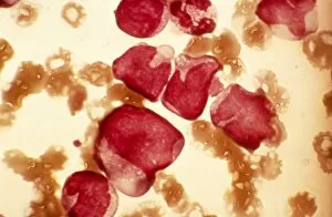
Background imageHistological Collection: Elliptocytosis, light micrograph

Elliptocytosis, light micrograph
Elliptocytosis. Light micrograph of red blood cells in a case of elliptocytosis. Red blood cells (erythrocytes) carry oxygen and carbon dioxide to and from body tissues

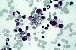
Background imageHistological Collection: Cystine in bone marrow, light micrograph

Cystine in bone marrow, light micrograph
Cystine in bone marrow. Light micrograph of crystals of cystine among blood cells in a sample of bone marrow. Cystine is an amino acid that can form crystals in urine

Background imageHistological Collection: Flu virus, TEM

Flu virus, TEM
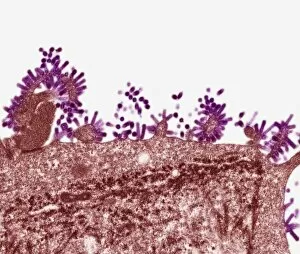
Flu virus. Transmission electron micrograph (TEM) of a section through influenza (flu) virus particles (virions, purple) budding from a host cell. This is the final stage in viral replication

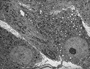
Background imageHistological Collection: Pancreatic exocrine cells, TEM

Pancreatic exocrine cells, TEM
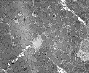
Pancreatic exocrine cells. Transmission electron micrograph (TEM) of a section through exocrine cells in the pancreas, showing numerous zymogen granules (circles), rough endoplasmic reticulum (ER)

Background imageHistological Collection: Bone marrow blood cells, light micrograph

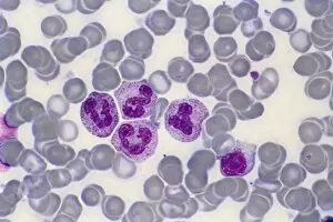
Bone marrow blood cells, light micrograph

Background imageHistological Collection: Glomerulus, TEM

Glomerulus, TEM
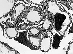
Glomerulus. Transmission electron micrograph (TEM) of a section through a nephron in a human kidney, showing a cluster of capillaries within a renal corpuscle, also known as a glomerulus

Background imageHistological Collection: Monocyte blood cell, light micrograph

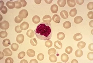
Monocyte blood cell, light micrograph. Monocytes are a type of white blood cell, part of the bodys immune system. Monocytes are the largest white blood cell


For sale as Licensed Images

Choose your image, Select your licence and Download the media

"Exploring the Intricacies of Histological Wonders: Unveiling the Hidden Beauty within Our Bodies" Step into the fascinating world of histology, where microscopic wonders come to life. Delve into Santiago Ramon Y Cajal's masterpiece, as his cortical grey matter schema unveils the intricate network that forms our brain's command center. Witness the mesmerizing complexity of a mammalian retina through a histological diagram, revealing how light is transformed into vision. Journey deeper into our cerebellum tissue, as a captivating light micrograph showcases its unique structure and function. Marvel at Ramon Y Cajal's genius once again as he unravels the secrets of synapse nerve junctions through an astonishing TEM image. Venture further into uncharted territory with a glimpse of hippocampus brain tissue, where memories are formed and stored. Observe Purkinje nerve cells in the cerebellum, their elegant arrangement hinting at their crucial role in coordinating movement. Witness nature's precision with kidney tubules in section; these delicate structures filter waste from our bloodstreams tirelessly. Explore human brain microscope slides and be awestruck by its intricate architecture - billions of neurons working harmoniously to shape who we are. Peer closely at rough endoplasmic reticulum through TEM imagery; this cellular powerhouse orchestrates protein synthesis within each living cell. Immerse yourself in glial stem cell culture under a light microscope - witness new life being nurtured for potential regeneration. Finally, marvel at the intricacy of brain tissue blood supply - countless vessels ensuring oxygen and nutrients reach every corner of this remarkable organ. Histological wonders unlock hidden beauty within us all – reminding us that even on a microscopic level, there is awe-inspiring complexity waiting to be discovered.