Histological Collection (#9)

"Exploring the Intricacies of Histological Wonders: Unveiling the Hidden Beauty within Our Bodies" Step into the fascinating world of histology

Background imageHistological Collection: Xavier Bichat, French pathologist

Xavier Bichat, French pathologist
Marie-Francois Xavier Bichat (1771-1802), French pathologist and founder of modern histology. Bichat worked at the Hotel-Dieu hospital in Paris

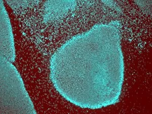
Background imageHistological Collection: Induced stem cells, light micrograph

Induced stem cells, light micrograph
Induced stem cells. Light micrograph of a cultured colony of induced human pluripotent stem (IPS) cells. Oct-4 (octamer-binding transcription factor 4) is cyan

Background imageHistological Collection: Induced nerve stem cells, micrograph

Induced nerve stem cells, micrograph
Induced nerve stem cells. Fluorescence light micrograph of neural (nerve) stem cells that have been created (induced) from human adult skin fibroblast cells by gene manipulation

Background imageHistological Collection: Stem cell-derived nerve cells

Stem cell-derived nerve cells. Fluorescence light micrograph of neural (nerve) stem cells that have been derived from human embryonic stem cells (HESC)

Background imageHistological Collection: Arteriole, TEM

Arteriole, TEM
Arteriole. Transmission electron micrograph (TEM) of a section through a small calibre arterial vessel called an arteriole

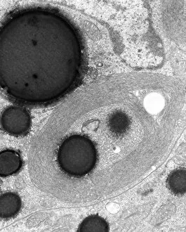
Background imageHistological Collection: Smooth endoplasmic reticulum, TEM

Smooth endoplasmic reticulum, TEM
Smooth endoplasmic reticulum. Transmission electron micrograph (TEM) showing smooth endoplasmic reticulum (ER, thin lines) inside a cell that is synthesising steroid hormones

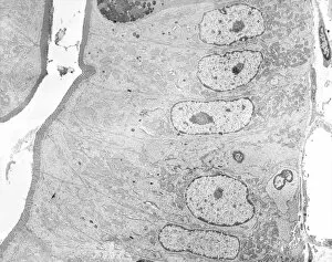
Background imageHistological Collection: Small bowel epithelium, TEM

Small bowel epithelium, TEM
Small bowel epithelium. Transmission electron micrograph (TEM) of a section through the epithelial lining of a villus in the small bowel, showing the enterocyte cells

Background imageHistological Collection: Lung tissue, confocal micrograph C014 / 4616

Lung tissue, confocal micrograph C014 / 4616
Lung tissue. Laser scanning confocal micrograph (LSCM) of a section through a human lung

Background imageHistological Collection: Kidney tissue, confocal micrograph C014 / 4611

Kidney tissue, confocal micrograph C014 / 4611
Kidney tissue. Laser scanning confocal micrograph (LSCM) of a section through a human kidney, showing the cell nuclei (dark)

Background imageHistological Collection: Liver portal triad, light micrograph C016 / 8490

Liver portal triad, light micrograph C016 / 8490
Liver portal triad. Fluorescence deconvolution micrograph of a section through a portal triad in liver tissue, showing hepatocyte cells (red)

Background imageHistological Collection: Liver portal triad, light micrograph C016 / 8489

Liver portal triad, light micrograph C016 / 8489
Liver portal triad. Fluorescence deconvolution micrograph of a section through a portal triad in liver tissue, showing hepatocyte cells (red)

Background imageHistological Collection: Liver portal triad, light micrograph C016 / 8488

Liver portal triad, light micrograph C016 / 8488
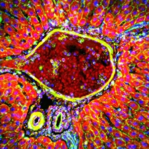
Liver portal triad. Fluorescence deconvolution micrograph of a section through a portal triad in liver tissue, showing hepatocyte cells (red)

Background imageHistological Collection: Kidney tissue, fluorescence micrograph C016 / 8484

Kidney tissue, fluorescence micrograph C016 / 8484
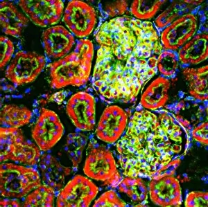
Kidney tissue. Fluorescence deconvolution micrograph of a section through a kidney, showing glomeruli (green), cell nuclei (blue dots), and renal tubules (red, circular)

Background imageHistological Collection: Heart muscle, fluorescence micrograph C016 / 8483

Heart muscle, fluorescence micrograph C016 / 8483
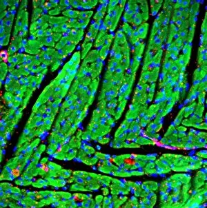
Heart muscle. Fluorescence deconvolution micrograph of a section through heart tissue, showing the angular distribution of the myocardium (cardiac muscle) fibres (green)

Background imageHistological Collection: Heart muscle, fluorescence micrograph C016 / 8481

Heart muscle, fluorescence micrograph C016 / 8481
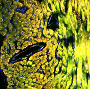
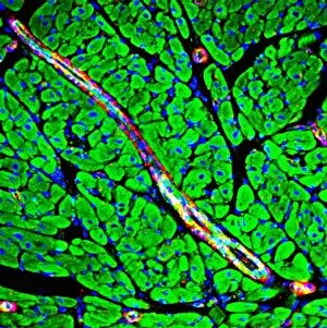
Heart muscle. Fluorescence deconvolution micrograph of a section through heart tissue, showing blood vessels (oval, centre-left and upper centre) running through the myocardium (cardiac muscle)

Background imageHistological Collection: Heart muscle, fluorescence micrograph C016 / 8478

Heart muscle, fluorescence micrograph C016 / 8478
Heart muscle. Fluorescence deconvolution micrograph of a section through heart tissue, showing a blood vessel (diagonal, centre) running through the myocardium (cardiac muscle, green)

Background imageHistological Collection: Heart muscle, fluorescence micrograph C016 / 8477

Heart muscle, fluorescence micrograph C016 / 8477
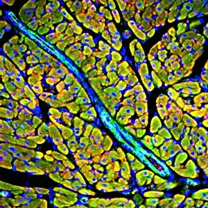
Heart muscle. Fluorescence deconvolution micrograph of a section through heart tissue, showing a blood vessel (blue, diagonal, centre) running through the myocardium (cardiac muscle, green)

Background imageHistological Collection: Adrenal gland, fluorescence micrograph C016 / 8472

Adrenal gland, fluorescence micrograph C016 / 8472
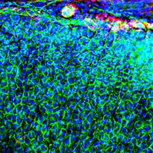
Adrenal gland. Fluorescence deconvolution micrograph of a section through an adrenal gland, showing the zona glomerulosa and zona fasciculata

Background imageHistological Collection: Kidney blood vessels, confocal micrograph C014 / 4610

Kidney blood vessels, confocal micrograph C014 / 4610
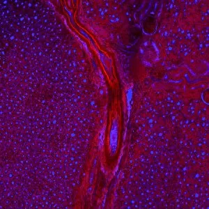
Kidney blood vessels. Laser scanning confocal micrograph (LSCM) of a section through a human kidney, showing a blood vessel (vertical, centre)

Background imageHistological Collection: Muscle tissue, light micrograph C014 / 4598

Muscle tissue, light micrograph C014 / 4598
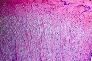
Muscle tissue. Light micrograph of a section through fibrous connective tissue from a longitudinal muscle tendon

Background imageHistological Collection: Arabidopsis thaliana embryo, micrograph C014 / 4590

Arabidopsis thaliana embryo, micrograph C014 / 4590
Arabidopsis thaliana embryo. Confocal light micrograph of a section through the embryo of a thale cress (Arabidopsis thaliana) plant

Background imageHistological Collection: Blood vessels, light micrograph C014 / 4562

Blood vessels, light micrograph C014 / 4562
Blood vessels. Light micrograph of a section through healthy human tissue showing an artery (large, red) and vein (small, red), surrounded by connective tissue (yellow)

Background imageHistological Collection: Brain nerve cells, TEM C014 / 0356

Brain nerve cells, TEM C014 / 0356
Brain nerve cells. Transmission electron micrograph (TEM) of a section through brain tissue from the cerebral cortex, showing numerous neurons (nerve cells) surrounded by axons and dendrites

Background imageHistological Collection: Brain cell, TEM C014 / 0358

Brain cell, TEM C014 / 0358
Brain cell. Transmission electron micrograph (TEM) of a section through an oligodendrocyte in human brain tissue. Oligodendrocytes occur in both the white

Background imageHistological Collection: Brain cell, TEM C014 / 0359

Brain cell, TEM C014 / 0359
Brain cell. Transmission electron micrograph (TEM) of a section through an oligodendrocyte in human brain tissue. Oligodendrocytes occur in both the white

Background imageHistological Collection: Lactating breast tissue, light micrograph C015 / 7102

Lactating breast tissue, light micrograph C015 / 7102
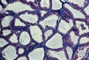
Lactating breast tissue. Light micrograph of a section through healthy breast (mammary gland) tissue during lactation. Glandular tissue in the breast has proliferated to produce spaces (alveoli)

Background imageHistological Collection: Healthy cervical smear, micrograph C015 / 6995

Healthy cervical smear, micrograph C015 / 6995
Healthy cervical smear. Light micrograph of healthy squamous epithelial cells from a smear test. A cervical smear, or pap test (after the papanicolaou used to stain the slides)

Background imageHistological Collection: Sebaceous gland, light micrograph C015 / 6911

Sebaceous gland, light micrograph C015 / 6911
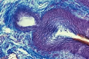
Sebaceous gland. Light micrograph of a section through breast tissue showing a sebaceous gland. These glands produce an oily protective substance called sebum

Background imageHistological Collection: Golgi apparatus, artwork C015 / 6764

Golgi apparatus, artwork C015 / 6764
Computer artwork of the Golgi apparatus of the human cell. This organelle functions as a central delivery system for the cell

Background imageHistological Collection: Breast tissue, light micrograph C015 / 6743

Breast tissue, light micrograph C015 / 6743
Breast tissue. Light micrograph of a section through healthy breast (mammary gland) tissue from a woman who is not pregnant

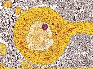
Background imageHistological Collection: Purkinje nerve cell, TEM C014 / 0582

Purkinje nerve cell, TEM C014 / 0582
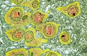
Purkinje nerve cell. Transmission electron micrograph (TEM) of a purkinje nerve cell (bright yellow, centre) from the cerebellum of the brain

Background imageHistological Collection: Canadian pondweed leaf, light micrograph

Canadian pondweed leaf, light micrograph
Canadian pondweed leaf. Light micrograph of a section through the leaf of a Canadian pondweed (Elodea canadensis) plant, showing the cells (squares)

Background imageHistological Collection: Corpus albicans, light micrograph C015 / 6226

Corpus albicans, light micrograph C015 / 6226
Corpus albicans. Light micrograph of a section through an ovary showing a corpus albicans (centre). Each menstrual cycle one egg (ovum) reaches maturity and develops into a Graafian follicle

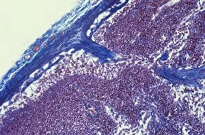
Background imageHistological Collection: Lymph node, light micrograph C015 / 6223

Lymph node, light micrograph C015 / 6223
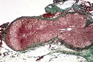
Lymph node. Light micrograph of a section through a lymph node. The blue tissue at right is the fibrous outer capsule of the node. Beneath it is the subcapsular sinus (white)

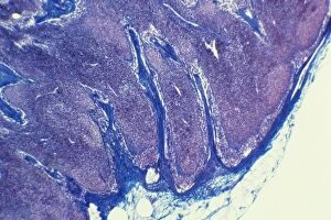
Background imageHistological Collection: Lymph node, light micrograph C015 / 6222

Lymph node, light micrograph C015 / 6222
Lymph node. Light micrograph of a section through a lymph node. The blue tissue at left is the fibrous outer capsule of the node. Beneath it is the subcapsular sinus (white)

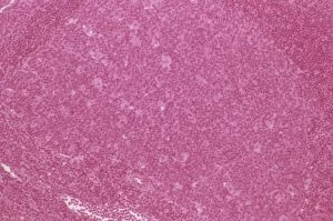
Background imageHistological Collection: Lymph node hyperplasia, light micrograph C015 / 6224

Lymph node hyperplasia, light micrograph C015 / 6224
Lymph node hyperplasia. Light micrograph of a section through a lymph node showing follicular hyperplasia. Hyperplasia is the abnormal proliferation of cells

Background imageHistological Collection: Pituitary gland C015 / 6420

Pituitary gland C015 / 6420
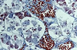
Pituitary gland. Light micrograph of a section through a healthy pituitary gland. The pituitary is a pea-sized organ found at the base of the brain

Background imageHistological Collection: Islet of Langerhans, TEM C015 / 6418

Islet of Langerhans, TEM C015 / 6418
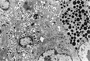
Islet of Langerhans. Transmission electron micrograph (TEM) of a section through cells clustered in an islet of Langerhans in the pancreas

Background imageHistological Collection: Adrenal cortex, light micrograph C015 / 6417

Adrenal cortex, light micrograph C015 / 6417
Adrenal cortex. Light micrograph of a section through a healthy adrenal cortex (red). The adrenal glands are found on top of the kidneys

Background imageHistological Collection: Sphagnum moss cells, light micrograph

Sphagnum moss cells, light micrograph
Sphagnum moss cells. Polarised light micrograph of a section through cells from Sphagnum sp. moss. Magnification: x400 when printed 10 centimetres wide

Background imageHistological Collection: Moss cells, light micrograph

Moss cells, light micrograph
Moss cells, polarised light micrograph. Magnification: x400 when printed 10 centimetres wide

Background imageHistological Collection: Magnolia stem, SEM C015 / 5128

Magnolia stem, SEM C015 / 5128
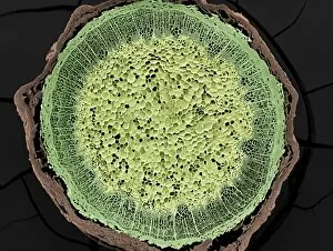
Magnolia stem. Coloured environmental scanning electron micrograph (ESEM) of a transverse section through the stem of a Magnolia sp. plant

Background imageHistological Collection: Magnolia stem, SEM C015 / 5124

Magnolia stem, SEM C015 / 5124
Magnolia stem. Coloured environmental scanning electron micrograph (ESEM) of a transverse section through the stem of a Magnolia sp. plant

Background imageHistological Collection: Intraglomerular mesangial cells, TEM C016 / 5837

Intraglomerular mesangial cells, TEM C016 / 5837
Intraglomerular mesangial cells. Transmission electron micrograph (TEM) of a section through intraglomerular mesangial cells from the glomerulus of a kidney

Background imageHistological Collection: Kidney glomerulus, TEM C016 / 5831

Kidney glomerulus, TEM C016 / 5831
Kidney glomerulus. Transmission electron micrograph (TEM) of a section through a renal glomerulus. At centre left is a capillary with a red blood cell (black) in its lumen

Background imageHistological Collection: Unmyelinated nerve, TEM C016 / 5805

Unmyelinated nerve, TEM C016 / 5805
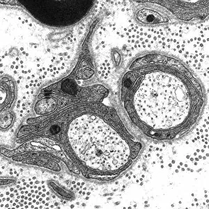
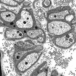
Unmyelinated nerve. Transmission electron micrograph (TEM) of a section through axon (nerve fibre) bundles of unmyelinated nerves

Background imageHistological Collection: Unmyelinated nerve, TEM C016 / 5804

Unmyelinated nerve, TEM C016 / 5804
Unmyelinated nerve. Transmission electron micrograph (TEM) of a section through axon (nerve fibre) bundles of unmyelinated nerves

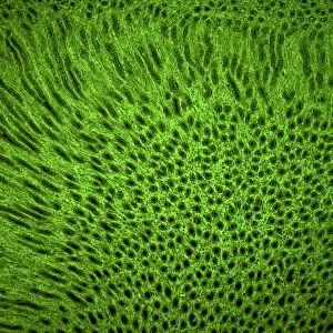
Background imageHistological Collection: Stomach wall, confocal light micrograph C016 / 3127

Stomach wall, confocal light micrograph C016 / 3127
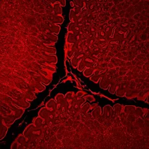
Stomach wall. Laser scanning confocal light micrograph of a section through the wall of a stomach


For sale as Licensed Images

Choose your image, Select your licence and Download the media

"Exploring the Intricacies of Histological Wonders: Unveiling the Hidden Beauty within Our Bodies" Step into the fascinating world of histology, where microscopic wonders come to life. Delve into Santiago Ramon Y Cajal's masterpiece, as his cortical grey matter schema unveils the intricate network that forms our brain's command center. Witness the mesmerizing complexity of a mammalian retina through a histological diagram, revealing how light is transformed into vision. Journey deeper into our cerebellum tissue, as a captivating light micrograph showcases its unique structure and function. Marvel at Ramon Y Cajal's genius once again as he unravels the secrets of synapse nerve junctions through an astonishing TEM image. Venture further into uncharted territory with a glimpse of hippocampus brain tissue, where memories are formed and stored. Observe Purkinje nerve cells in the cerebellum, their elegant arrangement hinting at their crucial role in coordinating movement. Witness nature's precision with kidney tubules in section; these delicate structures filter waste from our bloodstreams tirelessly. Explore human brain microscope slides and be awestruck by its intricate architecture - billions of neurons working harmoniously to shape who we are. Peer closely at rough endoplasmic reticulum through TEM imagery; this cellular powerhouse orchestrates protein synthesis within each living cell. Immerse yourself in glial stem cell culture under a light microscope - witness new life being nurtured for potential regeneration. Finally, marvel at the intricacy of brain tissue blood supply - countless vessels ensuring oxygen and nutrients reach every corner of this remarkable organ. Histological wonders unlock hidden beauty within us all – reminding us that even on a microscopic level, there is awe-inspiring complexity waiting to be discovered.