
Abdominal Aorta Collection
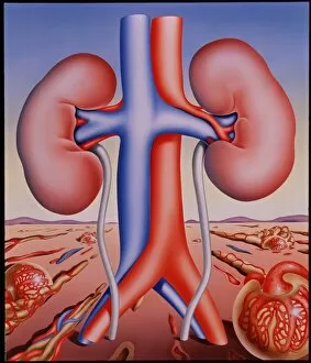
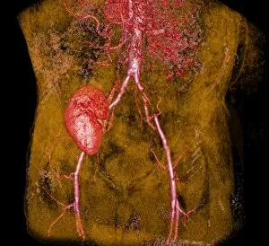
The abdominal aorta, depicted in this 1874 lithograph, is a vital component of the human bloodstream


For sale as Licensed Images
Choose your image, Select your licence and Download the media
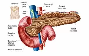
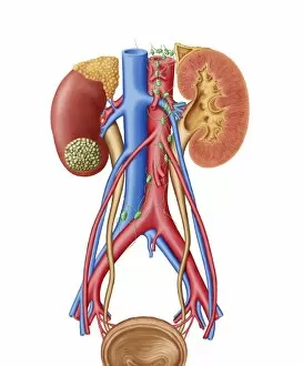
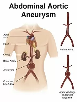
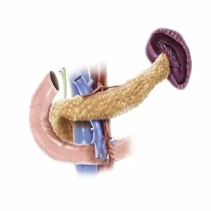
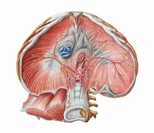
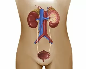
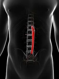
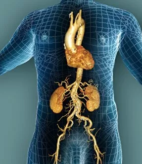
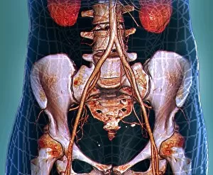
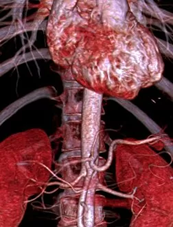
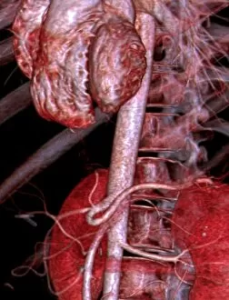
The abdominal aorta, depicted in this 1874 lithograph, is a vital component of the human bloodstream. Serving as the main artery that supplies oxygen-rich blood to various organs and tissues within the abdomen, it plays a crucial role in maintaining our overall health. Adjacent to the pancreas and kidneys, this large vessel can sometimes be affected by medical conditions such as kidney cancer or abdominal aortic aneurysm (AAA). In early stages of kidney cancer, tumors may become visible on the kidney itself, necessitating immediate attention for proper diagnosis and treatment. To better understand these conditions, medical illustrations like artist depictions of AAA with labels or detailed anatomical diagrams of the pancreas are invaluable resources. These visuals aid healthcare professionals in identifying abnormalities and guiding their interventions accordingly. Moreover, comprehending how different body systems interact is essential when considering diseases that affect multiple areas simultaneously. For instance, understanding both male and female urinary systems alongside blood pressure regulation through the circulatory system provides valuable insights into diagnosing complex issues involving these interconnected structures. Additionally, knowledge about related anatomy like the abdominal surface of diaphragm helps clinicians assess potential complications arising from disorders affecting adjacent regions. Ultimately, staying informed about our bodies' intricate workings empowers us to recognize signs and symptoms associated with specific ailments promptly. Medical charts displaying indicators of kidney cancer serve as useful references for individuals seeking information about potential warning signs they should be aware of. Exploring various aspects surrounding the abdominal aorta not only deepens our understanding but also promotes proactive healthcare practices aimed at early detection and effective management of related conditions.