Alga Collection (#14)

"Discovering the Intricate Beauty of Algae: From Ernst Haeckel's Art to 19th-Century Cyanotypes and SEM Images" Immerse yourself in the captivating world of algae

Background imageAlga Collection: Diatom shell, SEM

Diatom shell, SEM
Diatom shell. Coloured scanning electron micrograph (SEM) of the shell of a Biddulphia sp. diatom. The diatoms are a group of photosynthetic, single-celled algae containing about 10, 000 species

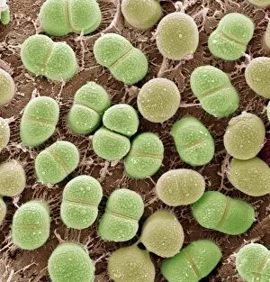
Background imageAlga Collection: Algae, SEM

Algae, SEM
Algae. Coloured scanning electron micrograph (SEM) of single-celled algae (green). Magnification: x4000 when printed 10 centimetres wide

Background imageAlga Collection: Coccoliths, SEM

Coccoliths, SEM
Coccoliths, coloured scanning electron micrograph (SEM). Coccoliths are individual plates of calcium carbonate formed by coccolithophores (single-celled algae)

Background imageAlga Collection: Eutrophication in a flooded field

Eutrophication in a flooded field. Muddy pool on a pathway running through a field of grazing cattle. The run-off of water contains urine and soluble dung and is ideal for the growth of algae

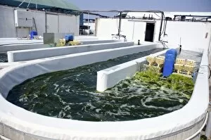
Background imageAlga Collection: Growing algae for fish food

Growing algae for fish food. Nannochloropsis sp. algae (green) being grown at a fishery. Once grown the algae is used to feed fish. Photographed at the Maagan Michael kibbutz, Coastal Plains, Israel

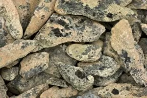
Background imageAlga Collection: Lichen-covered rocks

Lichen-covered rocks. Lichens on scree rocks on a mountainside. Lichens are organisms formed by the symbiotic association of a fungus and a green alga or cyanobacterium

Background imageAlga Collection: Whiteworm lichen (Thamnolia vermicularis)

Whiteworm lichen (Thamnolia vermicularis) on rocks. Lichens are organisms formed by the symbiotic association of a fungus and a green alga or cyanobacterium

Background imageAlga Collection: Algal colony, light micrograph

Algal colony, light micrograph. Colony of the green freshwater alga Coleochaete sentata. The whole plant is a disc composed of filaments packed close together, branching out from the centre

Background imageAlga Collection: Polluted river, USA

Polluted river, USA
Polluted river. This is a creek mouth on the Indian River, Titusville, Florida, USA

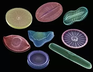
Background imageAlga Collection: Selection of diatoms, SEM

Selection of diatoms, SEM
Selection of diatoms, coloured scanning electron micrograph (SEM). Diatoms are single-celled photosynthetic algae, of which there are about 100, 000 species

Background imageAlga Collection: Spirulina cyanobacteria, light micrograph

Spirulina cyanobacteria, light micrograph
Spirulina cyanobacteria. Light micrograph of Spirulina platensis cyanobacteria (formally known as blue-green algae) filaments. Each filament is a colony of bacterial cells

Background imageAlga Collection: Coscinodiscus diatom, SEM

Coscinodiscus diatom, SEM
Coscinodiscus diatom. Coloured scanning electron micrograph (SEM) of a Coscinodiscus diatom (red). This is a marine diatom

Background imageAlga Collection: Skeleton shrimp and mussels

Skeleton shrimp and mussels
Skeleton shrimp (Caprella septentrionalis) and blue mussels (Mytilus edulis) on a brown algae (Laminaria sp.) frond

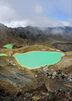
Background imageAlga Collection: Volcanic lakes, New Zealand

Volcanic lakes, New Zealand
Volcanic lakes. These are the Emerald Lakes in Tongariro National Park, on New Zealands North Island. The Tongariro National Park is the oldest national park in New Zealand

Background imageAlga Collection: Lace lichen (Ramalina menziesii)

Lace lichen (Ramalina menziesii) growing on the branches of a valley oak tree (Quercus lobata). Lichens are symbiotic organisms formed of a fungus and an alga that mutually benefit one another

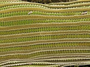
Background imageAlga Collection: Filamentous alga, SEM

Filamentous alga, SEM
Filamentous alga. Coloured scanning electron micrograph (SEM) of a filamentous green alga. It comprises rod-shaped cells, linked into filaments or strands

Background imageAlga Collection: Lichens

Lichens

Background imageAlga Collection: Desmid alga, SEM

Desmid alga, SEM
Desmid. Coloured scanning electron micrograph (SEM) of a desmid. Desmids are a common type of green algae and are usually found in freshwater

Background imageAlga Collection: Freshwater alga, light micrograph

Freshwater alga, light micrograph
Freshwater alga. Light micrograph of a freshwater alga, the water net Hydrodictyon, showing a full grown net (thick threads) and a young net still growing (thin threads)

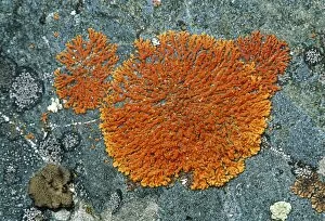
Background imageAlga Collection: Lichen (Teloschistes capensis)

Lichen (Teloschistes capensis). Lichens are symbiotic organisms formed of a fungus and an alga that mutually benefit one another. The alga is contained within the hyphae (filaments) of the fungus

Background imageAlga Collection: Spirogyra algae, light micrograph

Spirogyra algae, light micrograph
Spirogyra algae. Light micrograph of filaments from a Spirogyra sp. alga, showing its spiralling bands of chloroplasts (green). Central nuclei (round) are also seen in each cell

Background imageAlga Collection: Spirogyra algae

Spirogyra algae
Spirogyra algal strand. Confocal light micrograph of a filament of Spirogyra sp. green algae. This alga forms slimy threads called " water-silk" or " mermaids tresses" in stagnant

Background imageAlga Collection: Diatom assortment, SEMs

Diatom assortment, SEMs
Diatoms. Coloured scanning electron micrographs (SEMs) of a circular arrangement of various diatoms. Diatoms are planktonic unicellular algae


For sale as Licensed Images

Choose your image, Select your licence and Download the media

"Discovering the Intricate Beauty of Algae: From Ernst Haeckel's Art to 19th-Century Cyanotypes and SEM Images" Immerse yourself in the captivating world of algae, as we delve into its mesmerizing artistry and scientific wonders. Ernst Haeckel, a renowned biologist and artist, showcases the delicate intricacy of diatom algae through his stunning illustrations. These masterpieces reveal the symmetrical beauty hidden within these microscopic organisms. Step back in time with pressed seaweed specimens C016 / 6127 from centuries ago. Preserved meticulously, they offer a glimpse into the diverse forms and colors that algae can take on. The cyanotype technique used during the 19th century captures their essence in ethereal blue hues, adding an enchanting touch to their already fascinating nature. Calcareous phytoplankton observed under scanning electron microscopy (SEM) unravels another layer of algal marvels. Discosphaera tubifera, a coccolithophore species adorned with intricate calcium carbonate plates, showcases nature's architectural genius at work. Dictyota dichotoma emerges as a striking example of brown algae thriving along coastlines worldwide. Its branching structure creates an artistic spectacle underwater while providing essential habitats for marine life. Diatoms continue to amaze us with their geometric patterns when viewed under SEM imaging techniques. Their microscopic beauty is unveiled through meticulous detailing that reveals their remarkable diversity and ecological significance. Venturing further into aquatic realms, Fucus bulbosus or kelp stands tall as one of nature's giants beneath ocean waves. This majestic alga plays a crucial role in coastal ecosystems by offering shelter to numerous marine organisms. Pond life surprises us with its vibrant assortment of microorganisms where diatoms once again steal the show under SEM imaging techniques. Their intricate designs resemble miniature works of art floating amidst water bodies' tranquility.