
Alimentary Canal Collection

"Journey Through the Alimentary Canal

Background imageAlimentary Canal Collection: Human digestive system, artwork

Human digestive system, artwork. At top, food from the mouth forms a bolus that is swallowed down the oesophagus. At centre, the bolus is about to enter the stomach (arrow)

Background imageAlimentary Canal Collection: Intestinal microvilli, TEM

Intestinal microvilli, TEM
Intestinal microvilli, coloured transmission electron micrograph (TEM). The microvilli are finger-like projections (seen here in circular cross-section)

Background imageAlimentary Canal Collection: Vintage Anatomical Print of the Human Alimentary Canal, 1912 (screen print)

Vintage Anatomical Print of the Human Alimentary Canal, 1912 (screen print)
3034945 Vintage Anatomical Print of the Human Alimentary Canal, 1912 (screen print) by American School, (20th century); (add.info)

Background imageAlimentary Canal Collection: Diagram showing the Alimentary Canal, which passes right through the body, c1934

Diagram showing the Alimentary Canal, which passes right through the body, c1934. From The Treasury of Knowledge. [Richard Clay & Sons, Ltd. Bungay, Suffolk, 1934]

Background imageAlimentary Canal Collection: Intestinal villi, SEM

Intestinal villi, SEM
Intestinal villi. Coloured scanning electron micrograph (SEM) of a section through a part of the small intestine, showing the villi that cover its inner surface

Background imageAlimentary Canal Collection: Severe gastritis, endoscope view C016 / 3898

Severe gastritis, endoscope view C016 / 3898
Severe gastritis, endoscope view. Gastritis is inflammation of the stomach lining. It causes pain on eating or drinking, and may cause nausea and vomiting

Background imageAlimentary Canal Collection: Human intestines, artwork

Human intestines, artwork
Human intestines. Artwork of a human torso with the intestines highlighted in colour. They consist of the small intestine (pink), circled by the large intestine (light brown)

Background imageAlimentary Canal Collection: Small intestine, light micrograph

Small intestine, light micrograph
Small intestine. Light micrograph of a section through the small intestine. The finger-like projections at left are the villi. These increase the surface area for the absorption of food

Background imageAlimentary Canal Collection: Ulcerative pancolitis C018 / 0928

Ulcerative pancolitis C018 / 0928
Ulcerative pancolitis. Endoscope view of the appendix aperture in the caecum (start of the large intestine) of an 11 year old patient with ulcerative pancolitis showing inflamed mucosa

Background imageAlimentary Canal Collection: Healthy large intestine, 3D CT scan C016 / 6509

Healthy large intestine, 3D CT scan C016 / 6509
Healthy large intestine. Coloured 3D computed tomography (CT) scan of the abdomen of a 37 year old female patient showing a healthy large intestine

Background imageAlimentary Canal Collection: Healthy large intestine, 3D CT scan C016 / 6508

Healthy large intestine, 3D CT scan C016 / 6508
Healthy large intestine. Coloured 3D computed tomography (CT) scan of the abdomen of a 37 year old female patient showing a healthy large intestine

Background imageAlimentary Canal Collection: Total mesorectal excision, artwork C016 / 6996

Total mesorectal excision, artwork C016 / 6996
Total mesorectal excision (TME). Artwork of the pelvis and the area of the colon and rectum that is removed in total mesorectal excision

Background imageAlimentary Canal Collection: Healthy large intestine, 3D CT scan C016 / 6507

Healthy large intestine, 3D CT scan C016 / 6507
Healthy large intestine. Coloured 3D computed tomography (CT) scan of the abdomen of a 37 year old female patient showing a healthy large intestine

Background imageAlimentary Canal Collection: Oesophagus, artwork

Oesophagus, artwork
Oesophagus. Computer artwork of the oesophagus, which carries food from the mouth to the stomach

Background imageAlimentary Canal Collection: Pancreatic cancer, artwork C016 / 8735

Pancreatic cancer, artwork C016 / 8735
Pancreatic cancer. Artwork of part of the digestive system, showing a carcinoma (white, lower right) of the head of the pancreas (light brown)

Background imageAlimentary Canal Collection: Gastrointestinal disorders, artwork C016 / 8734

Gastrointestinal disorders, artwork C016 / 8734
Gastrointestinal disorders. Artwork of part of the digestive system, showing ulcers on the internal lining of the stomach (centre right) and the internal lining of the duodenum (centre left)

Background imageAlimentary Canal Collection: Healthy stomach, X-ray

Healthy stomach, X-ray
Healthy stomach, coloured X-ray. The stomach is a curved, sac-like organ that receives food from the oesophagus and passes it to the small intestine

Background imageAlimentary Canal Collection: Bowel disease in the colon C016 / 8338

Bowel disease in the colon C016 / 8338
Bowel disease in the colon. Endoscopic view of diverticular disease (diverticulosis) affecting the sigmoid colon. Diverticulosis causes sacs (diverticula)

Background imageAlimentary Canal Collection: Diverticulitis C017 / 8015

Diverticulitis C017 / 8015
Diverticulitis. X-ray of the colon of a patient with diverticulitis. A diverticulum is a small pouch-like structure that protrudes out of weak spots in the intestinal wall

Background imageAlimentary Canal Collection: Diverticulitis C017 / 8014

Diverticulitis C017 / 8014
Diverticulitis. X-ray of the colon of a patient with diverticulitis. A diverticulum is a small pouch-like structure that protrudes out of weak spots in the intestinal wall

Background imageAlimentary Canal Collection: Diverticulitis C017 / 7835

Diverticulitis C017 / 7835
Diverticulitis. X-ray of the colon of a patient with diverticulitis. A diverticulum is a small pouch-like structure that protrudes out of weak spots in the intestinal wall

Background imageAlimentary Canal Collection: Swallowed marble, X-ray C017 / 7748

Swallowed marble, X-ray C017 / 7748
Swallowed marble. X-ray of the abdomen of a 4 year old male patient that has swallowed a marble

Background imageAlimentary Canal Collection: Swallowed marble, X-ray C017 / 7749

Swallowed marble, X-ray C017 / 7749
Swallowed marble. X-ray of the abdomen of a 4 year old male patient that has swallowed a marble

Background imageAlimentary Canal Collection: Stomach anatomy, artwork C016 / 2871

Stomach anatomy, artwork C016 / 2871
Stomach anatomy, artwork. The stomach is the organ that receives food from the oesophagus (gullet, top) and passes it onwards to the intestines

Background imageAlimentary Canal Collection: Stomach anatomy, artwork C016 / 2858

Stomach anatomy, artwork C016 / 2858
Stomach anatomy. Artwork showing the position of the stomach (red) in relation to other abdominal organs and the human skeleton, in an oblique right-frontal view from below

Background imageAlimentary Canal Collection: Stomach anatomy, artwork C016 / 2857

Stomach anatomy, artwork C016 / 2857
Stomach anatomy. Artwork showing the position of the stomach (red) in relation to other abdominal organs and the human skeleton, as seen in an oblique left-frontal view

Background imageAlimentary Canal Collection: Stomach anatomy, artwork C016 / 2856

Stomach anatomy, artwork C016 / 2856
Stomach anatomy. Artwork showing the position of the stomach (red) in relation to other abdominal organs and the human skeleton, as seen in an anterior (frontal) view

Background imageAlimentary Canal Collection: Stomach anatomy, artwork C016 / 2855

Stomach anatomy, artwork C016 / 2855
Stomach anatomy. Artwork showing the position of the stomach (red) in relation to other abdominal organs and the human skeleton, as seen in an anterior (frontal) view

Background imageAlimentary Canal Collection: Stomach, artwork C014 / 0001

Stomach, artwork C014 / 0001
Stomach. Computer artwork of a human stomach. At top is the oesophagus (tube), which carries food from the mouth to the stomach (centre)

Background imageAlimentary Canal Collection: Hover fly gut, SEM

Hover fly gut, SEM
Hover fly gut. Coloured scanning electron micrograph (SEM) of the gut of a hover fly (family Syrphidae). The hover fly feeds on pollen and nectar. Magnification unknown

Background imageAlimentary Canal Collection: Duodenal microvilli

Duodenal microvilli
Microvilli in duodenum. Coloured transmission electron micrograph (TEM) of a section through the human duodenum, showing microvilli on the surface epithelium (lining)

Background imageAlimentary Canal Collection: Small intestine villus, SEM

Small intestine villus, SEM
Small intestine villus. Coloured scanning electron micrograph (SEM) of a freeze fracture section through a villus from the mucosal lining of the small intestine

Background imageAlimentary Canal Collection: Secretory cells in pancreas, SEM

Secretory cells in pancreas, SEM
Pancreatic secretory cells. Coloured scanning electron micrograph (SEM) of a freeze-fracture through a healthy pancreas, showing the secretory tissue

Background imageAlimentary Canal Collection: Small intestine lining, SEM

Small intestine lining, SEM
Small intestine villi. Coloured scanning electron micrograph (SEM) of villi (folds) on the lining of the small intestine. Villi greatly increase the intestinal surface area for absorbing nutrients

Background imageAlimentary Canal Collection: Small intestine villi, SEM

Small intestine villi, SEM
Small intestine villi. Coloured scanning electron micrograph (SEM) of villi (folds) on the lining of the small intestine. Villi greatly increase the intestinal surface area for absorbing nutrients

Background imageAlimentary Canal Collection: Duodenum secretory cells

Duodenum secretory cells. Coloured transmission electron micrograph (TEM) of a section through the human duodenum, showing secretory cells of the surface epithelium (lining)

Background imageAlimentary Canal Collection: Stomach wall, SEM

Stomach wall, SEM
Stomach wall. Coloured scanning electron micrograph (SEM) of a section through a freeze- fractured fold in the stomach wall

Background imageAlimentary Canal Collection: Stomach, computer artwork

Stomach, computer artwork
Stomach. Computer artwork of the stomach, the first major organ of the digestive system. Food enters the stomach from the oesophagus (upper centre)

Background imageAlimentary Canal Collection: Oesophagus, X-ray

Oesophagus, X-ray
Oesophagus, coloured X-ray. The oesophagus (orange) is the tube that carries food and drink from the mouth to the stomach. It has been visualised here using a barium swallow

Background imageAlimentary Canal Collection: Oesophagus, SEM

Oesophagus, SEM
Oesophagus. Coloured scanning electron micrograph (SEM) of a freeze-fracture through the muscularis externa of the oesophagus. This forms part of the muscularis mucosa found throughout the gut

Background imageAlimentary Canal Collection: Oesophagus epithelium, SEM

Oesophagus epithelium, SEM
Oesophagus epithelium. Coloured scanning electron micrograph (SEM) of the lining of the human oesophagus or gullet (yellow) and two unknown bacteria (pink & purple)

Background imageAlimentary Canal Collection: Internal anatomy, 16th century diagram

Internal anatomy, 16th century diagram
Internal anatomy. 16th century anatomical diagram of the internal organs of the human torso. The diagram, known as a situs figure, is from Magnus Hundts Antropologium de hominis dignitate (Leipzig)

Background imageAlimentary Canal Collection: Internal anatomy, 15th century diagram

Internal anatomy, 15th century diagram
Internal anatomy. 15th century anatomical diagram of the internal organs of the human head and torso. The diagram, known as a situs figure

Background imageAlimentary Canal Collection: Wound sites, 15th century artwork

Wound sites, 15th century artwork
Wound sites. 15th century anatomical diagram illustrating the various injuries and their common sites. The diagram, known as a wound man, is from Hieronymus Brunschwigs Book of Cirurgia (Strassburg)

Background imageAlimentary Canal Collection: Irritable bowel syndrome

Irritable bowel syndrome. Artwork of a digestive system suffering from irritable bowel syndrome (IBS). IBS is also known as spastic colon or irritable colon syndrome

Background imageAlimentary Canal Collection: Crohns disease, X-ray

Crohns disease, X-ray

Background imageAlimentary Canal Collection: Ulcerative colitis, SEM

Ulcerative colitis, SEM
Ulcerative colitis. Coloured scanning electron micrograph (SEM) of freeze-fractured bowel tissue from a patient suffering from ulcerative colitis


For sale as Licensed Images

Choose your image, Select your licence and Download the media

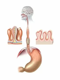
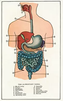
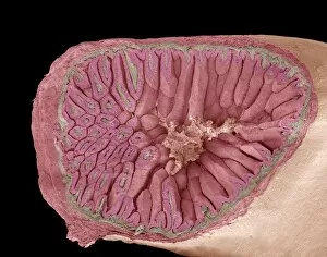
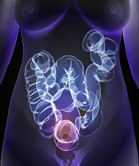
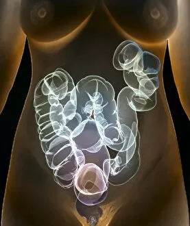
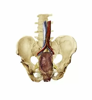
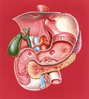
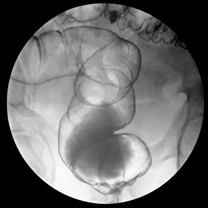
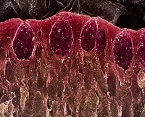
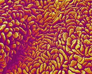
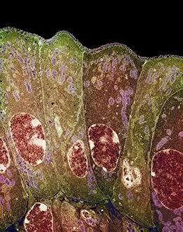
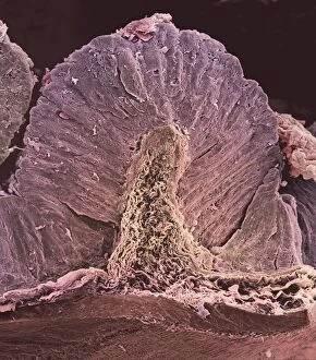
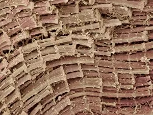
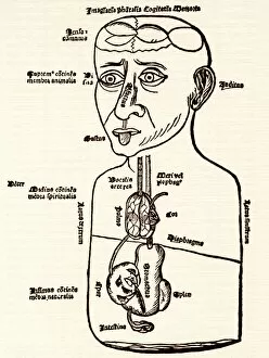
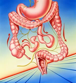
"Journey Through the Alimentary Canal: Exploring the Marvels of Our Digestive System" Step into the fascinating world of the human digestive system as we embark on a visual exploration. From vintage anatomical prints to intricate artwork, witness the beauty and complexity of our alimentary canal. Intriguingly detailed, an artistic representation from 1912 showcases the entirety of this remarkable system. The diagram vividly illustrates how it passes right through our bodies, highlighting its crucial role in nourishing us. Zooming in further, electron microscopy reveals intestinal microvilli with astonishing clarity. These tiny structures increase surface area for nutrient absorption, emphasizing nature's ingenious design. Moving forward in time, a captivating light micrograph captures the intricacies of the small intestine. Its vibrant colors and delicate textures remind us of its vital function in digestion and nutrient extraction. However, not all is smooth sailing within this wondrous canal. Medical images shed light on conditions such as ulcerative pancolitis and colon cancer locations depicted through thought-provoking artwork. These reminders serve as a call to action for maintaining good health and seeking early detection. But fear not. A healthy large intestine emerges from a 3D CT scan like an architectural marvel—a testament to resilience and well-being. Such scans provide invaluable insights into our internal workings while inspiring awe at their precision. Finally, we encounter total mesorectal excision—an artful representation that highlights surgical interventions aimed at treating specific ailments within this complex network. It serves as a reminder that medical advancements continue to evolve alongside our understanding of this incredible system. The alimentary canal—more than just a pathway for food—is an intricate masterpiece crafted by evolution itself. With each image encountered along this journey, we gain deeper appreciation for its wonders and are reminded to nurture it with care.