
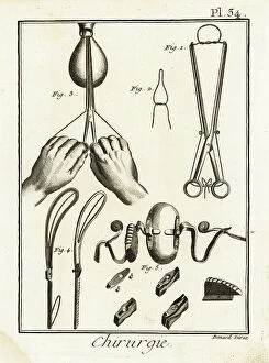
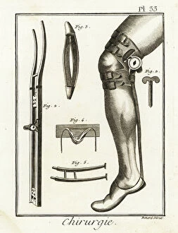
Anatomical Illustration Collection
"Exploring the Intricacies of Anatomy: A Journey through Anatomical Illustration" Step into the world of anatomical illustration


For sale as Licensed Images
Choose your image, Select your licence and Download the media
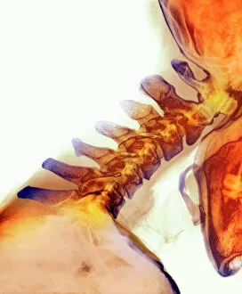
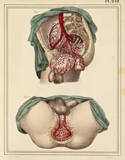
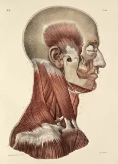
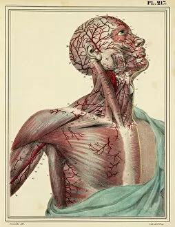
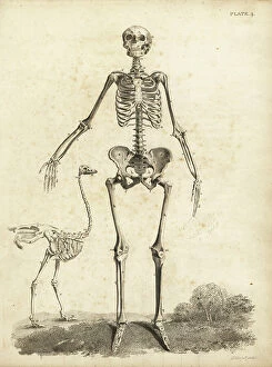
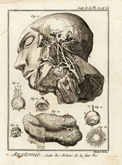
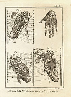
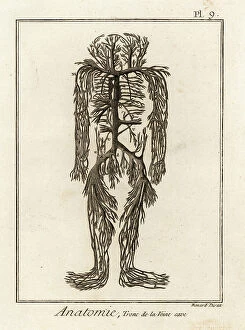
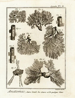
"Exploring the Intricacies of Anatomy: A Journey through Anatomical Illustration" Step into the world of anatomical illustration, where intricate details and scientific precision come to life. From extended neck vertebrae to mesmerizing X-ray images, this captivating art form unveils the hidden wonders within our bodies. Travel back in time to 1825 as you witness a masterpiece depicting male groin arteries, showcasing the delicate network that sustains life. Marvel at the 1831 artwork revealing head and neck muscles, intricately sketched with meticulous accuracy. Embark on a visual expedition through history with an exploration of head and chest arteries from another stunning 1825 artwork. Witness how these vital pathways intertwine like lifelines connecting every part of our being. Delve deeper into facial anatomy as you encounter face and neck muscles meticulously illustrated in an awe-inspiring 1831 artwork. Each stroke reveals the complexity beneath our skin, reminding us of nature's exquisite design. Uncover the secrets held by cervical spinal nerves portrayed in a remarkable 1844 artwork. These intricate pathways carry messages throughout our body, orchestrating movement and sensation with astonishing precision. Prepare for a chilling revelation as an anatomical view of a tooth exposes its demonic source behind toothaches—a haunting depiction reminiscent of hell itself. This macabre yet fascinating representation reminds us of both beauty and pain coexisting within us. Witness medical advancements brought to life through computer-generated imagery portraying angioplasty—an innovative technique revolutionizing cardiovascular care. Immerse yourself in this digital marvel that merges science with artistry seamlessly. Journey further into hand muscle anatomy showcased in an enchanting 1831 artwork—each sinewy line capturing strength and dexterity essential for human interaction. Appreciate how these intricate structures enable us to create, touch, and connect with one another. Explore forearm muscles depicted in yet another breathtaking piece from 1831—their interplay and strength showcased in a symphony of lines.