
Anatomist Collection (#10)
Anatomists have played a crucial role in unraveling the mysteries of the human body throughout history


For sale as Licensed Images
Choose your image, Select your licence and Download the media
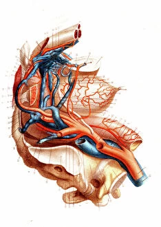
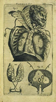
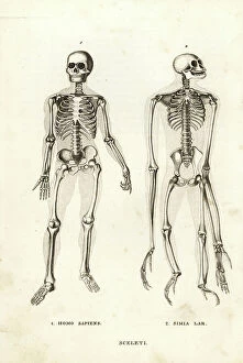
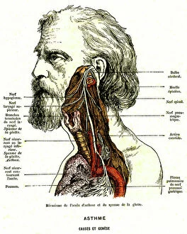
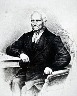
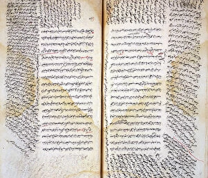
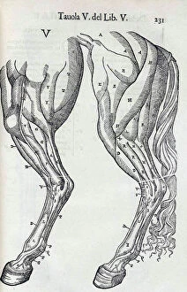
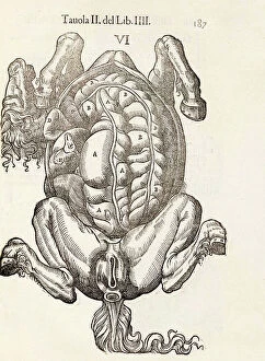
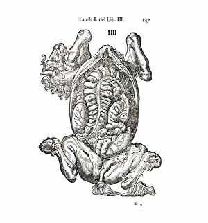
Anatomists have played a crucial role in unraveling the mysteries of the human body throughout history. One such pioneer was Robert Burton, whose extensive research and writings on anatomy left an indelible mark on the field. Another notable figure was EUSTACHIO Bartolomeo, an Italian anatomist who made significant contributions during the 16th century. Intriguingly, a lithograph titled "Odin questioning Mimir" captures the essence of curiosity that drives anatomists to seek knowledge. This thirst for understanding is exemplified by Sir Richard Owen, a renowned British anatomist from the 19th century whose work continues to inspire modern scientists. Richard Owen's dedication to his craft is evident in various depictions of him throughout history. In one caricature from Vanity Fair in 1873, aptly titled "Old bones, " he is portrayed as a distinguished scholar with wisdom etched onto his face. An image from his study at BMNH in 1883 showcases his meticulous approach towards dissecting and studying specimens. The intricate details captured in engravings like "The Sacred Heart of Jesus" or "The pollen of plants" demonstrate how anatomists explore not only human anatomy but also delve into other aspects of nature's design. Richard Owen himself had a profound impact on paleontology, as depicted in C016 / 5008 - an image that pays homage to his groundbreaking discoveries. Throughout history, heart anatomists have held a special place within this scientific realm. Figures like Guillaume Dupuytren and William Hunter dedicated their lives to unraveling the complexities of this vital organ. Their contributions paved the way for advancements in cardiac medicine that continue to save lives today. From ancient times until now, these brilliant minds have shaped our understanding of ourselves and our world through their tireless pursuit of knowledge about our bodies' inner workings. The legacy they leave behind serves as an inspiration for future generations aspiring to unravel the secrets of human anatomy.