
Anatomy Collection (#2)
"Unveiling the Intricacies of Anatomy: From Sensory Homunculus to Motor Homunculus" Step into the fascinating world of anatomy, where every detail tells a story


For sale as Licensed Images
Choose your image, Select your licence and Download the media
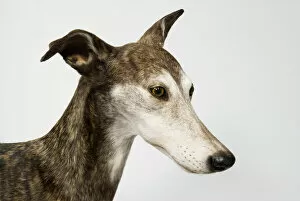
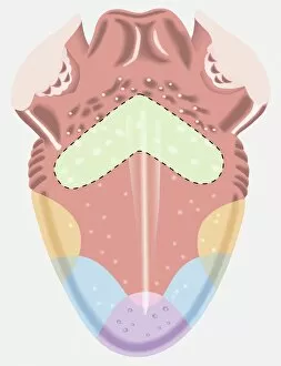
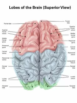
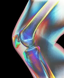
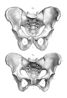
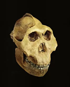
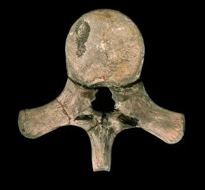
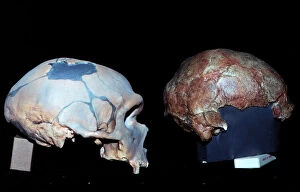
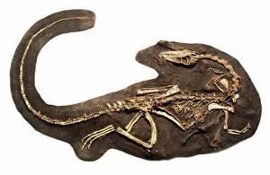
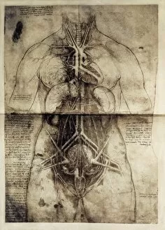
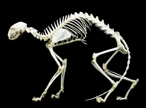
"Unveiling the Intricacies of Anatomy: From Sensory Homunculus to Motor Homunculus" Step into the fascinating world of anatomy, where every detail tells a story. Explore the depths of human and animal structures as we delve into this captivating subject. Let's begin with Leonardo da Vinci's masterpiece, "Head of a Bear. " Created in 1480 but discovered in 1945, this artwork showcases da Vinci's unparalleled ability to capture anatomical accuracy. The intricate details reveal his keen observation skills and deep understanding of form. Moving on to our furry friends, the greyhound takes center stage. Discovering the anatomy of these majestic creatures unveils their remarkable speed and agility. From their sleek skeleton to their muscular build, it is evident why they are renowned for their racing abilities. But what about us humans? Enter the sensory homunculus – a visual representation that depicts how our brain perceives different body parts based on sensitivity levels. This map reveals intriguing insights into how our senses are distributed throughout our bodies. X-ray images provide another dimension to understanding anatomy. Take a glimpse at normal knees through an X-ray lens – marvel at the complexity hidden beneath our skin while appreciating its delicate balance between strength and flexibility. The backbone is often considered one of nature's most ingenious designs. Our human backbone includes ribs and pelvis, providing stability while allowing movement – truly an architectural marvel worth exploring further. Venturing deeper within ourselves, let us explore the intricacies of the human brain from an inferior view. Witness its complex network of connections responsible for controlling various bodily functions - truly awe-inspiring. As we journey back in time, we encounter hominid crania – remnants that shed light on our evolutionary history. These ancient skulls offer glimpses into early forms such as Australopithecus afarensis (AL 288-1), famously known as Lucy - bridging gaps between past and present.