
Angiogenesis Collection
Angiogenesis, the process of blood vessel formation, plays a crucial role in various pathological conditions


For sale as Licensed Images
Choose your image, Select your licence and Download the media
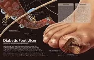
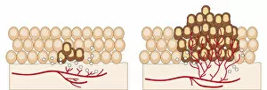
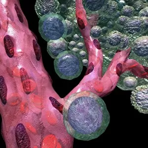
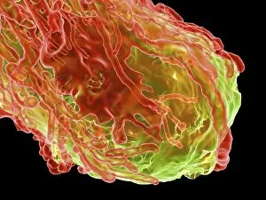
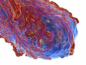
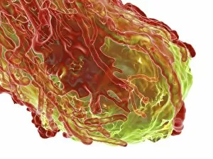
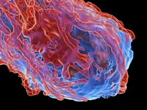
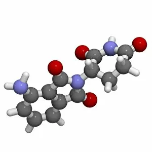
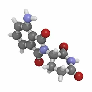
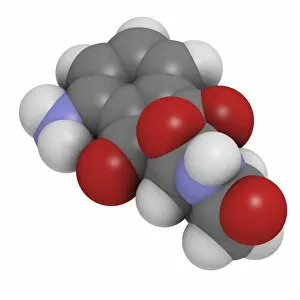
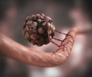
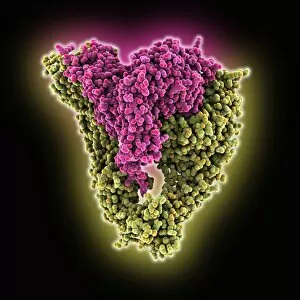
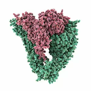
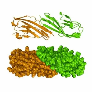
Angiogenesis, the process of blood vessel formation, plays a crucial role in various pathological conditions. In histopathology and the pathophysiology of diabetic foot ulcers, often impaired, leading to delayed wound healing and increased risk of infection. Through digital illustrations depicting the angiogenesis process, we can observe how dormant tumors invade surrounding tissues and grow into malignant masses. These visual representations provide valuable insights into the mechanisms behind tumor progression. Cross-sectional biomedical illustrations further shed light on how tumors obtain nutrients through angiogenesis. By developing an intricate network of blood vessels, tumors ensure their survival and growth by tapping into the body's resources. Artwork showcasing blood vessel formation emphasizes the importance in sustaining life processes. The intricate patterns formed by these vessels highlight their critical role in delivering oxygen and nutrients to different organs and tissues. Computer-generated images portraying tumors serve as powerful visuals that depict the destructive nature of cancer cells. These artworks aim to raise awareness about this devastating disease while highlighting its dependence on angiogenesis for sustenance. In addition to exploring tumor biology, medical illustrations also feature drugs like Regorafenib and Pomalidomide used in colorectal cancer treatment. These medications target specific pathways involved in tumor-induced angiogenesis, offering hope for patients fighting against this relentless disease. Overall, these captivating visuals not only showcase the complexity but also emphasize its significance in both normal physiological processes and pathological conditions such as cancer development. Understanding this intricate process opens doors for innovative therapeutic strategies aimed at inhibiting or promoting angiogenesis based on specific medical needs.