
Antibodies Collection
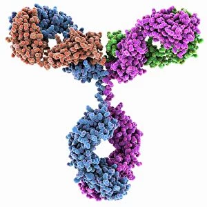
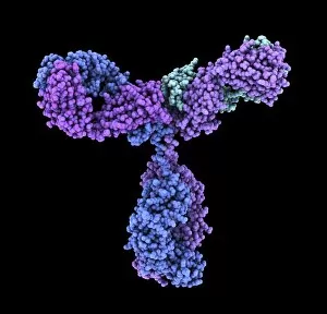
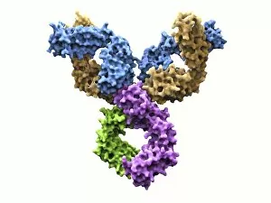
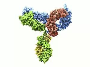
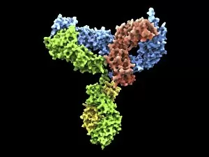
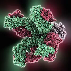
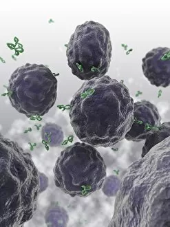
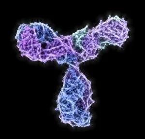
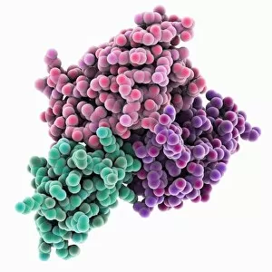
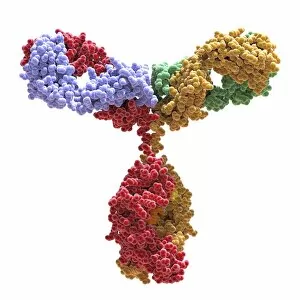
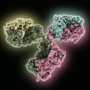
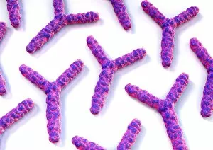
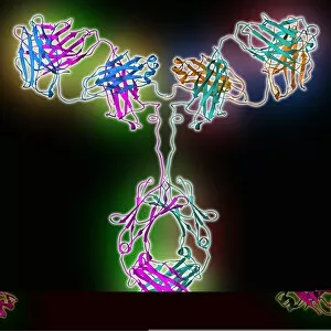
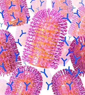
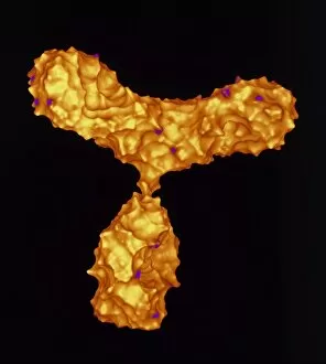
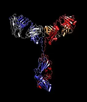
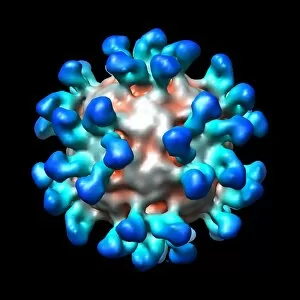
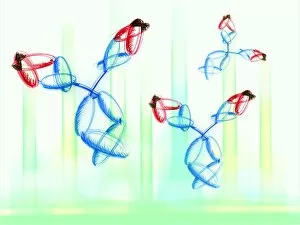
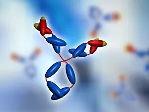
"Unleashing the Power of Antibodies: The Guardians of our Immune System" Immunoglobulin G antibody molecule F007 / 9894, a remarkable defender against pathogens


For sale as Licensed Images
Choose your image, Select your licence and Download the media
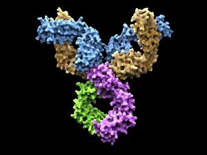
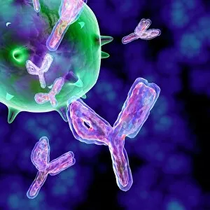
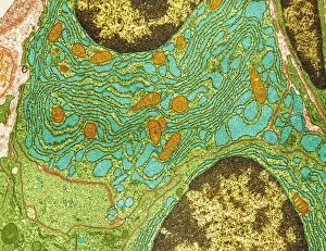
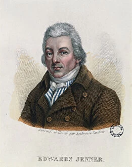
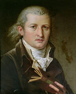
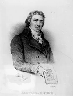
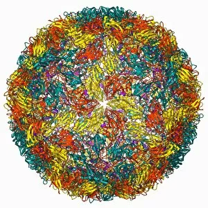
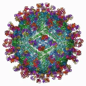
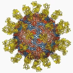
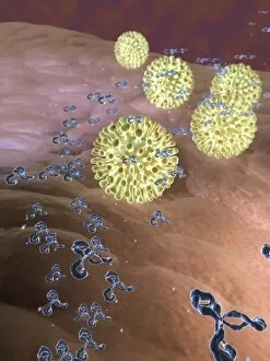
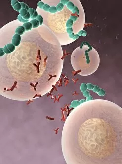
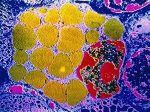
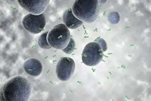
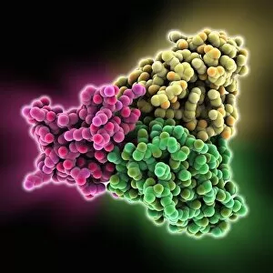
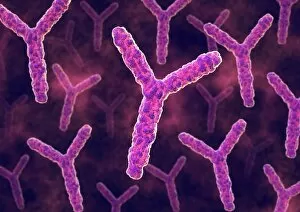
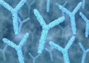
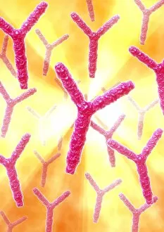
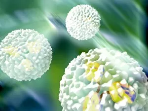
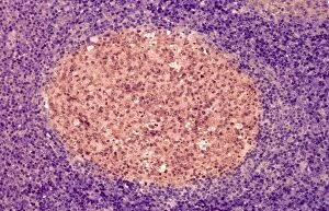
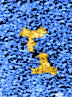
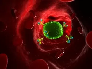
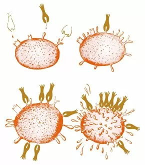
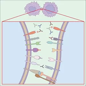
"Unleashing the Power of Antibodies: The Guardians of our Immune System" Immunoglobulin G antibody molecule F007 / 9894, a remarkable defender against pathogens, stands tall in this captivating artwork. These they can the unsung heroes that protect us from harmful invaders. Plasma cells, captured through a TEM image, tirelessly produce Immunoglobulin G antibody molecules to combat infections and maintain our well-being. Their intricate structure enables them to recognize specific antigens and neutralize threats with precision. Edward Jenner (1749-1823), depicted in various forms - coloured engraving, oil on canvas, lithography by de Frey - pioneered vaccination using cowpox virus. His groundbreaking work laid the foundation for harnessing antibodies' potential to prevent diseases. Intriguingly diverse yet unified in purpose, Immunoglobulin G antibody molecules emerge as an army defending our bodies against foot-and-mouth disease virus F006 / 9556 and countless other pathogens. They bind to these invaders like lock and key, rendering them harmless. These extraordinary proteins play a crucial role in immunology by recognizing foreign substances and triggering immune responses. Through their unique binding abilities, they mark intruders for destruction while sparing healthy cells. As we delve into the world of antibodies, we unveil their immense importance in safeguarding human health. From Edward Jenner's pioneering efforts to cutting-edge research today, these tiny warriors continue to shape medical advancements and save lives worldwide. Let us celebrate the awe-inspiring power – nature's guardians standing strong against adversity.