
Arthrosis Collection
"Arthrosis: A Closer Look at Joint Degeneration" Arthrosis, also known as osteoarthritis, is a degenerative joint disease that affects millions of people worldwide


For sale as Licensed Images
Choose your image, Select your licence and Download the media
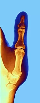
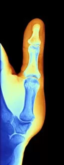
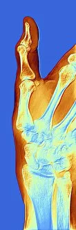
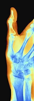
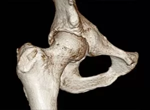
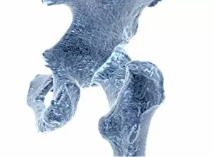
"Arthrosis: A Closer Look at Joint Degeneration" Arthrosis, also known as osteoarthritis, is a degenerative joint disease that affects millions of people worldwide. This condition can cause pain, stiffness, and reduced mobility in various joints of the body. One common area where arthrosis occurs is in the hand. X-ray images such as F006 / 4616, F006 / 4605, F006 / 4595, and F006 / 4598 reveal the telltale signs of arthritic changes within these delicate joints. The bony growths and narrowed joint spaces seen on these x-rays are indicative of this chronic condition. However, it's not just the hands that suffer from arthrosis. Other weight-bearing joints like the knee and hip can also be affected. Detailed imaging techniques like MRI provide a clearer picture of bone damage caused by this disease. The three-dimensional bone reconstructions from MRI data (C016 / 4694, C016 / 4614, C016 / 4611, C016/4607) showcase how arthrosis impacts these crucial joints. CT scans are another valuable tool for diagnosing joint diseases like arthrosis. By capturing detailed cross-sectional images of affected areas, they help healthcare professionals assess the extent of damage caused by this condition. Understanding the molecular aspects behind arthrosis is equally important for developing effective treatments. One molecule called Syndecan-4 has been found to play a role in cartilage breakdown associated with this disease process. While there is no cure for arthrosis currently available, early detection through diagnostic imaging allows for timely intervention strategies to manage symptoms effectively. Physical therapy exercises tailored to each patient's needs along with medication options can alleviate pain and improve quality of life.