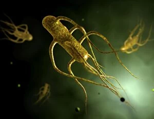
Bacteria Collection (#5)

"Bacteria: The Microscopic Marvels of Yellowstone National Park" In the heart of Wyoming, USA lies the breathtaking Yellowstone National Park

Background imageBacteria Collection: Nitrobacter sp bacteria, TEM

Nitrobacter sp bacteria, TEM

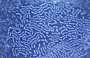
Background imageBacteria Collection: A colony of bacteria seen under a microscope

A colony of bacteria seen under a microscope

Background imageBacteria Collection: Lazzaro Spallanzani, Italian biologist, 1874

Lazzaro Spallanzani, Italian biologist, 1874. Spallanzani (1729-1799) worked on bacteria, disproving spontaneous generation, on digestion, where he was first to use the term gastric juice

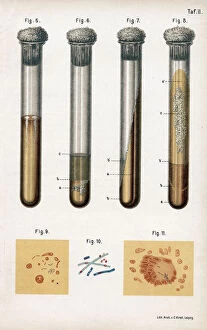
Background imageBacteria Collection: Various types of bacteria, 1889

Various types of bacteria, 1889. 5: pneumonia culture; 6: albumen from a rotten egg; 7& 8: tuberculosis as prepared by Koch; 9: sputum from a TB patient; 10: anthrax bacillus; 11

Background imageBacteria Collection: Essence of Parliament, 1883. Artist: Harry Furniss

Essence of Parliament, 1883. Artist: Harry Furniss
Essence of Parliament, 1883. Cartoon marking the opening of the first session of Parliament for 1883, showing the politicians as microbes in a drop of water

Background imageBacteria Collection: bacteria

bacteria
microscopic image

Background imageBacteria Collection: Fumigating a Room

Fumigating a Room
This young woman covers her mouth to protect herself from breathing in harmful bacteria as she seals a doorway and keyhole of a room in her house for fumigation from disease. Date: early 1930s

Background imageBacteria Collection: Advertisement for Salutaris bottled table water

Advertisement for Salutaris bottled table water, pure, distilled and aerated -- no foreign bodies permitted. Date: early 20th century

Background imageBacteria Collection: Pattern in Canary Spring, Yellowstone National Park, Montana / Wyoming

Pattern in Canary Spring, Yellowstone National Park, Montana / Wyoming
Pattern in Canary Spring, Yellowstone National Park (Montana, Wyoming)

Background imageBacteria Collection: A diagram showing aspergilloma affecting human lung

A diagram showing aspergilloma affecting human lung

Background imageBacteria Collection: Cross section biomedical illustration on grid of Worm, Fungi, Protozoa

Cross section biomedical illustration on grid of Worm, Fungi, Protozoa, and Bacteria infection and infestation

Background imageBacteria Collection: Picture No. 11675606

Picture No. 11675606
Patterns of Paenibacillus bacteria on petri dish - T type (tip-splitting morphotype). Date:

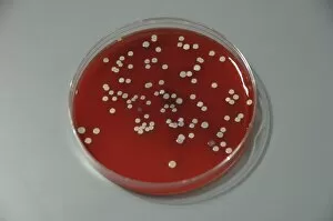
Background imageBacteria Collection: Red blood cells on an agar plate are used to diagnose infection

Red blood cells on an agar plate are used to diagnose infection. The plate on the left shows a positive staphyloccus infection

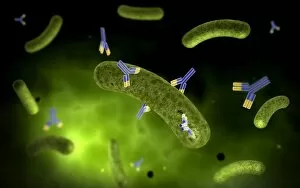
Background imageBacteria Collection: Conceptual image of antibody attaching and killing bacteria

Conceptual image of antibody attaching and killing bacteria

Background imageBacteria Collection: Microscopic view of histoplasmosis

Microscopic view of histoplasmosis. Histoplasmosis is an infection in the lungs caused by inhaling the spores of a fungus. This fungus, called Histoplasma capsulatum

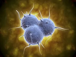
Background imageBacteria Collection: Microscopic view of diplococcus bacterium

Microscopic view of diplococcus bacterium

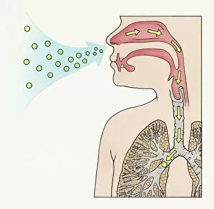
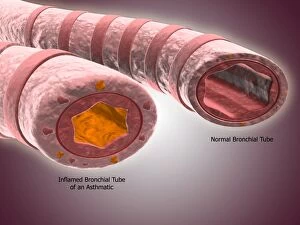
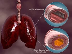
Background imageBacteria Collection: Trachea cross-section showing normal and asthmatic bronchiole

Trachea cross-section showing normal and asthmatic bronchiole
Trachea cross-section showing comparison of normal and asthmatic bronchiole

Background imageBacteria Collection: Conceptual image of cholerae bacteria

Conceptual image of cholerae bacteria

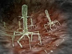
Background imageBacteria Collection: Microscopic view of bacteriophages on the surface of a bacteria

Microscopic view of bacteriophages on the surface of a bacteria

Background imageBacteria Collection: Electron micrograph of negative-stained Prochlorococcus

Electron micrograph of negative-stained Prochlorococcus. A cyanophage is a virus that infects cyanobacteria
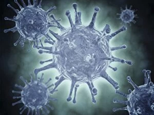
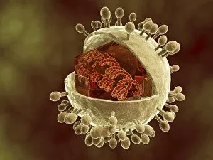
Background imageBacteria Collection: Conceptual image of the Hepatitis C virus

Conceptual image of the Hepatitis C virus. Hepatitis C virus is a small, enveloped, positive-sense single-stranded RNA virus of the family Flaviviridae

Background imageBacteria Collection: Development of fistula

Development of fistula

Background imageBacteria Collection: Lower gums with braces and plaque on teeth

Lower gums with braces and plaque on teeth

Background imageBacteria Collection: Trachea cross-section with lungs showing normal and asthmatic bronchiole

Trachea cross-section with lungs showing normal and asthmatic bronchiole
Trachea cross-section with lungs, showing comparison of normal and asthmatic bronchiole

Background imageBacteria Collection: Microscopic view of Methicillin-resistant Staphylococcus aureus

Microscopic view of Methicillin-resistant Staphylococcus aureus (MRSA). MRSA is a bacterium responsible for several difficult-to-treat infections in humans

Background imageBacteria Collection: Conceptual image of RNA virus replication

Conceptual image of RNA virus replication

Background imageBacteria Collection: Microscopic view of E. coli bacteria cells

Microscopic view of E. coli bacteria cells
Microscopic view of Escherichia coli bacteria cells, commonly known as E. Coli. E. coli is a common type of bacteria that can get into food, such as beef and vegetables

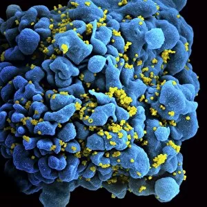
Background imageBacteria Collection: Colorized image of HIV-infected H9 T-cell

Colorized image of HIV-infected H9 T-cell

Background imageBacteria Collection: Bacteria from human skin grown on agar in the laboratory

Bacteria from human skin grown on agar in the laboratory

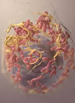
Background imageBacteria Collection: 3D structure of melanoma cell

3D structure of melanoma cell
3D structure of a melanoma cell derived by ion abrasion scanning electron microscopy

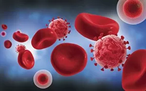
Background imageBacteria Collection: Microscopic view of blood cells with virus

Microscopic view of blood cells with virus

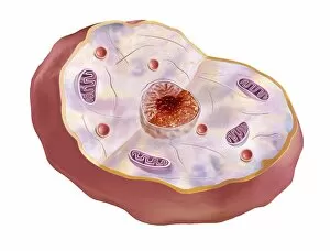
Background imageBacteria Collection: Human cell anatomy

Human cell anatomy

Background imageBacteria Collection: Three stages of periodontal disease

Three stages of periodontal disease

Background imageBacteria Collection: Image of a petri plate used to culture cells

Image of a petri plate used to culture cells

Background imageBacteria Collection: Microscopic view of the capsid protein of Nudaurelia capensis omega virus

Microscopic view of the capsid protein of Nudaurelia capensis omega virus

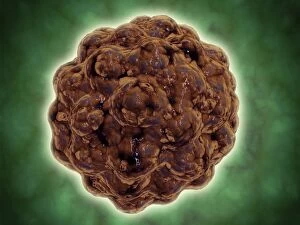
Background imageBacteria Collection: Conceptual image of a viral spore

Conceptual image of a viral spore

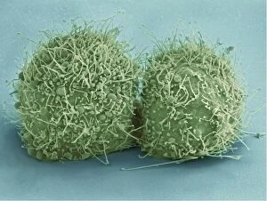
Background imageBacteria Collection: Scanning electron micrograph of just-divided HeLa cells

Scanning electron micrograph of just-divided HeLa cells. Zeiss Merlin HR-SEM

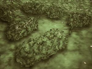
Background imageBacteria Collection: Microscopic view of Bacilli bacteria

Microscopic view of Bacilli bacteria

Background imageBacteria Collection: Microscopic view of Anthrax

Microscopic view of Anthrax. Anthrax is an acute disease caused by the bacterium Bacillus anthracis. Most forms of the disease are lethal, and it affects both humans and animals

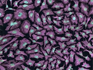
Background imageBacteria Collection: Multiphoton fluorescence image of HeLa cells

Multiphoton fluorescence image of HeLa cells with cytoskeletal microtubules (magenta) and DNA (cyan)

Background imageBacteria Collection: Microscopic view of streptococcus

Microscopic view of streptococcus

Background imageBacteria Collection: A colorized scanning electron micrograph of a white blood cell eating MRSA

A colorized scanning electron micrograph of a white blood cell eating MRSA
A colorized scanning electron micrograph of a white blood cell eating an antibiotic resistant strain of Staphylococcus aureus bacteria, commonly known as MRSA

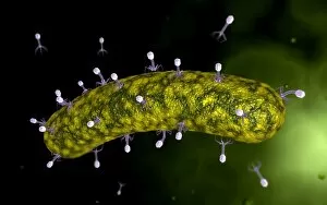
Background imageBacteria Collection: Microscopic view of bacteriophage attacking bacteria

Microscopic view of bacteriophage attacking bacteria

Background imageBacteria Collection: Conceptual image of Lactobacillus acidophilus

Conceptual image of Lactobacillus acidophilus. Lactobacillus is a gram-positive bacteria that occurs naturally in the human and animal gastrointestinal tract and mouth

Background imageBacteria Collection: Conceptual image of human cytomegalovirus

Conceptual image of human cytomegalovirus. Cytomegalovirus is a genus of the viral family Herpesviridae

Background imageBacteria Collection: Microscopic view of cocci bacterium

Microscopic view of cocci bacterium

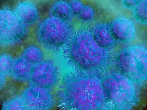
Background imageBacteria Collection: Conceptual image of salmonella typhi causing typhoid

Conceptual image of salmonella typhi causing typhoid


For sale as Licensed Images

Choose your image, Select your licence and Download the media

"Bacteria: The Microscopic Marvels of Yellowstone National Park" In the heart of Wyoming, USA lies the breathtaking Yellowstone National Park, a haven for nature enthusiasts and scientists alike. Amidst its natural wonders, one can find an extraordinary display of microbial life at the Grand Prismatic Spring in the Midway Geyser Basin. Picture No. 11675590 captures this mesmerizing sight, showcasing vibrant hues that seem to dance upon the water's surface. However, what may appear as an artistic masterpiece is actually a thriving community of bacteria. Zooming in closer with SEM C018 / 8596 reveals a neutrophil engulfing MRSA - Methicillin-resistant Staphylococcus aureus. This image serves as a reminder of how these tiny organisms can pose significant threats to human health. Another SEM image showcases E. Coli bacteria, reminding us that not all it can harmful; some play crucial roles in our digestive system and even aid scientific research. Moving back to Yellowstone's enchantment, we encounter Salmonella bacteria through another captivating SEM photograph. These microscopic creatures serve as a stark reminder that even within such serene landscapes, dangers lurk unseen. Coloured TEM imagery introduces us to Yersinia pestis bacteria – responsible for causing devastating outbreaks like the infamous Black Death throughout history. Such visuals emphasize their intricate structures and highlight why understanding them is vital for public health efforts. Venturing beyond real-life images into computer artwork brings forth a beta DNA segment surrounded by spheres—a representation of genetic material essential for bacterial survival and reproduction. Artistic depictions also shed light on various cell types found within these microorganisms' complex world—each playing unique roles necessary for their survival and proliferation. Historical diagrams reveal cultures of Anthrax—an infectious disease with severe consequences if left unchecked—underscoring humanity's ongoing battle against bacterial infections throughout time. Tuberculosis bacteria make their appearance too—a haunting reminder that despite medical advancements, infectious diseases continue to challenge us.