
Bones Collection (page 4)

"Bones: Unveiling the Hidden Structures of Life" Delving into the depths of human anatomy

Background imageBones Collection: Frontal View of Human Skull, 1917 (lithograph)

Frontal View of Human Skull, 1917 (lithograph)
2622467 Frontal View of Human Skull, 1917 (lithograph) by German School, (20th century); Private Collection; (add.info.: Vintage illustration of an anterior or frontal view of an anatomically correct)

Background imageBones Collection: Anatomical View of the Human Torso, 1914 (engraving)

Anatomical View of the Human Torso, 1914 (engraving)
2622096 Anatomical View of the Human Torso, 1914 (engraving) by German School, (20th century); Private Collection; (add.info.: Vintage anatomical study of the human torso, frontal view)

Background imageBones Collection: Common Raven (Corvus corax) Skull, Probably Cornwall, England

Common Raven (Corvus corax) Skull, Probably Cornwall, England
The Common Raven is the largest member of the Crow Family and is one of the most widely distributed birds worldwide. It is all black in colour with a long, wedge shaped tail

Background imageBones Collection: Neon Halloween Skull

Neon Halloween Skull
Sarah Manovski

Background imageBones Collection: Face Paint Creativity

Face Paint Creativity
Tori E Bohn

Background imageBones Collection: Huli Fortune Teller

Huli Fortune Teller
Tori E Bohn

Background imageBones Collection: Fish-skin helmet, c.1800-1900 (fish skin)

Fish-skin helmet, c.1800-1900 (fish skin)
7264988 Fish-skin helmet, c.1800-1900 (fish skin); Manchester Museum, The University of Manchester, England; (add.info.: Helmet made from a large Porcupine fish)

Background imageBones Collection: Whalebone fid, 1850-1900 (whalebone)

Whalebone fid, 1850-1900 (whalebone)
7339928 Whalebone fid, 1850-1900 (whalebone) by Unknown Artist, (19th century); 28x3.2 cm; National Maritime Museum, London

Background imageBones Collection: Bishops Crosier of walrus tusk

Bishops Crosier of walrus tusk
5304704 Bishops Crosier of walrus tusk.; (add.info.: Bishops crosier (staff) of walrus tusk, found with the bones of Bishop Pali Jonsson of Skalholt (d)

Background imageBones Collection: 'The Caffre Man.' Native of South Africa holding three spears, naked

"The Caffre Man." Native of South Africa holding three spears, naked
5856471 "The Caffre Man." Native of South Africa holding three spears, naked, but wearing a necklace of bones and a red belt around his waist.; (add.info)

Background imageBones Collection: A seam rubber, a sailor's tool used in the manufacture of sails, early 19th century (whalebone)

A seam rubber, a sailor's tool used in the manufacture of sails, early 19th century (whalebone)
MMM5476242 A seam rubber, a sailor's tool used in the manufacture of sails, early 19th century (whalebone); National Maritime Museum, London, UK; (add.info.: A seam rubber)

Background imageBones Collection: A seam rubber, a sailor's tool used in the manufacture of sails, early 19th century (whalebone)

A seam rubber, a sailor's tool used in the manufacture of sails, early 19th century (whalebone)
MMM5476232 A seam rubber, a sailor's tool used in the manufacture of sails, early 19th century (whalebone); National Maritime Museum, London, UK; (add.info.: A seam rubber)

Background imageBones Collection: How to carve ham (b/w photo)

How to carve ham (b/w photo)
8657629 How to carve ham (b/w photo) by English Photographer, (20th century); Private Collection; (add.info.: How to carve ham)

Background imageBones Collection: Skeleton of a crocodile, Crocodylus niloticus. Copperplate engraving by Heath after an

Skeleton of a crocodile, Crocodylus niloticus. Copperplate engraving by Heath after an illustration by George Shaw
FLO4694870 Skeleton of a crocodile, Crocodylus niloticus. Copperplate engraving by Heath after an illustration by George Shaw from his General Zoology, Amphibia, London

Background imageBones Collection: Anatomy of the human skeleton, 1839 (lithograph)

Anatomy of the human skeleton, 1839 (lithograph)
FLO4684794 Anatomy of the human skeleton, 1839 (lithograph); (add.info.: Anatomy of the human skeleton. Lithograph from Lorenz Oken's Universal Natural History)

Background imageBones Collection: Wilhelm Rontgen

Wilhelm Rontgen
3733270 Wilhelm Rontgen.; (add.info.: Photograph of Wilhelm Rontgen (1845-1923) a German mechanical engineer and physicist

Background imageBones Collection: Human female skeleton from the front, with ostrich skeleton by William Cheselden

Human female skeleton from the front, with ostrich skeleton by William Cheselden
FLO4565814 Human female skeleton from the front, with ostrich skeleton by William Cheselden. Copperplate engraving by Edward Mitchell after an anatomical illustration by Jean-Joseph Sue from John

Background imageBones Collection: Longitudinal sections of the femur 1, 2 and of the tibia 3. Copperplate engraving by Edward

Longitudinal sections of the femur 1, 2 and of the tibia 3. Copperplate engraving by Edward Mitchell after an
FLO4565745 Longitudinal sections of the femur 1, 2 and of the tibia 3. Copperplate engraving by Edward Mitchell after an anatomical illustration by Jean-Joseph Sue from John Barclay's A Series of

Background imageBones Collection: Views of the femur and rotula bones in the human leg. Copperplate engraving by Edward Mitchell

Views of the femur and rotula bones in the human leg. Copperplate engraving by Edward Mitchell after an anatomical
FLO4565729 Views of the femur and rotula bones in the human leg. Copperplate engraving by Edward Mitchell after an anatomical illustration by Jean-Joseph Sue from John Barclay's A Series of

Background imageBones Collection: Views of the tibia bones in the human leg. Copperplate engraving by Edward Mitchell after an

Views of the tibia bones in the human leg. Copperplate engraving by Edward Mitchell after an anatomical illustration by
FLO4565727 Views of the tibia bones in the human leg. Copperplate engraving by Edward Mitchell after an anatomical illustration by Jean-Joseph Sue from John Barclay's A Series of Engravings of

Background imageBones Collection: Bones of a primitive horse

Bones of a primitive horse
3507366 Bones of a primitive horse; (add.info.: Bones of a primitive horse dating back 10, 000 years.); Universal History Archive

Background imageBones Collection: Illustration depicting the uncovering of the prehistoric past

Illustration depicting the uncovering of the prehistoric past
3522058 Illustration depicting the uncovering of the prehistoric past; (add.info.: Illustration depicting the uncovering of the prehistoric past. Dated 19th Century); Universal History Archive/UIG

Background imageBones Collection: Skull of an early homo sapiens

Skull of an early homo sapiens
3507255 Skull of an early homo sapiens; (add.info.: Skull of an early homo sapiens found in Eastern Europe.); Universal History Archive

Background imageBones Collection: Comb and trial-piece

Comb and trial-piece
3330290 Comb and trial-piece; (add.info.: Comb and trial-piece made from carved bone and antler. Dated 9th Century.); Universal History Archive/UIG

Background imageBones Collection: Anatomical illustration

Anatomical illustration
2625713 Anatomical illustration; (add.info.: Anatomical drawing by William Cheselden in Osteographia, or The anatomy of the bones. (London: 1733).); Universal History Archive/UIG

Background imageBones Collection: Anatomical illustration

Anatomical illustration
2625710 Anatomical illustration; (add.info.: Anatomical drawing by William Cheselden in Osteographia, or The anatomy of the bones. (London: 1733).); Universal History Archive/UIG

Background imageBones Collection: Anatomical illustration

Anatomical illustration
2625709 Anatomical illustration; (add.info.: Anatomical drawing by William Cheselden in Osteographia, or The anatomy of the bones. (London: 1733).); Universal History Archive/UIG

Background imageBones Collection: Anatomical illustration

Anatomical illustration
2625706 Anatomical illustration; (add.info.: Anatomical drawing by William Cheselden in Osteographia, or The anatomy of the bones. (London: 1733).); Universal History Archive/UIG

Background imageBones Collection: Skeleton of a European perch, Perca fluviatilis, from Dr. Barclay's Museum, Edinburgh

Skeleton of a European perch, Perca fluviatilis, from Dr. Barclay's Museum, Edinburgh
FLO4565818 Skeleton of a European perch, Perca fluviatilis, from Dr. Barclay's Museum, Edinburgh. Copperplate engraving by Edward Mitchell after an anatomical illustration by Robert Kaye Greville

Background imageBones Collection: Rear view of the human trunk skeleton including spine, ribs and pelvis

Rear view of the human trunk skeleton including spine, ribs and pelvis. Copperplate engraving by Edward Mitchell after
FLO4565792 Rear view of the human trunk skeleton including spine, ribs and pelvis. Copperplate engraving by Edward Mitchell after an anatomical illustration by Jean-Joseph Sue from John Barclay's

Background imageBones Collection: Views of the bones in the human foot from ankle to toe. Copperplate engraving by Edward Mitchell

Views of the bones in the human foot from ankle to toe. Copperplate engraving by Edward Mitchell after an anatomical
FLO4565738 Views of the bones in the human foot from ankle to toe. Copperplate engraving by Edward Mitchell after an anatomical illustration by Jean-Joseph Sue from John Barclay's A Series of

Background imageBones Collection: Bones of the carpus, metacarpus and fingers in the human hand

Bones of the carpus, metacarpus and fingers in the human hand. Copperplate engraving by Edward Mitchell after an
FLO4565722 Bones of the carpus, metacarpus and fingers in the human hand. Copperplate engraving by Edward Mitchell after an anatomical illustration from John Barclay's A Series of Engravings of

Background imageBones Collection: Sternum and cartilage of the human ribcage, with several ribs

Sternum and cartilage of the human ribcage, with several ribs. Copperplate engraving by Edward Mitchell after an
FLO4565702 Sternum and cartilage of the human ribcage, with several ribs. Copperplate engraving by Edward Mitchell after an anatomical illustration by Jean-Joseph Sue from John Barclay's A Series

Background imageBones Collection: Ribs and sternum in the human ribcage. Copperplate engraving by Edward Mitchell after an

Ribs and sternum in the human ribcage. Copperplate engraving by Edward Mitchell after an anatomical illustration by
FLO4565699 Ribs and sternum in the human ribcage. Copperplate engraving by Edward Mitchell after an anatomical illustration by Jean-Joseph Sue from John Barclay's A Series of Engravings of

Background imageBones Collection: Front view of the human trunk skeleton including spine, ribs and pelvis

Front view of the human trunk skeleton including spine, ribs and pelvis. Copperplate engraving by Edward Mitchell after
FLO4565690 Front view of the human trunk skeleton including spine, ribs and pelvis. Copperplate engraving by Edward Mitchell after an anatomical illustration by Jean-Joseph Suefrom John Barclay's

Background imageBones Collection: The application of Liston's Long Splint

The application of Liston's Long Splint
6116637 The application of Liston's Long Splint, ; (add.info.: The application of Liston's Long Splint. Long leg splinting is used to stabilize injuries by decreasing movement)

Background imageBones Collection: Reduction of dislocated thigh using pulleys. From The Household Physician

Reduction of dislocated thigh using pulleys. From The Household Physician, published c.1898 (engraving)
6116647 Reduction of dislocated thigh using pulleys. From The Household Physician, published c.1898 (engraving); Photo © Ken Welsh. All rights reserved 2024

Background imageBones Collection: Anatomical illustration

Anatomical illustration
2625712 Anatomical illustration; (add.info.: Anatomical drawing by William Cheselden in Osteographia, or The anatomy of the bones. (London: 1733).); Universal History Archive/UIG

Background imageBones Collection: Anatomical illustration

Anatomical illustration
2625711 Anatomical illustration; (add.info.: Anatomical drawing by William Cheselden in Osteographia, or The anatomy of the bones. (London: 1733).); Universal History Archive/UIG

Background imageBones Collection: Scrimshaw whale tooth 'The Sailor Boy', mid 19th century (whale tooth)

Scrimshaw whale tooth "The Sailor Boy", mid 19th century (whale tooth)
7339166 Scrimshaw whale tooth "The Sailor Boy", mid 19th century (whale tooth) by Unknown Artist, (19th century); 8x23x8.9 cm; National Maritime Museum, London

Background imageBones Collection: Whale tooth, 1830-40 (whale tooth)

Whale tooth, 1830-40 (whale tooth)
7339922 Whale tooth, 1830-40 (whale tooth) by Unknown Artist, (19th century); 4x6.5x16 cm; National Maritime Museum, London

Background imageBones Collection: Engraving depicting the bones of the human foot, 19th century

Engraving depicting the bones of the human foot, 19th century
5309455 Engraving depicting the bones of the human foot, 19th century; (add.info.: Engraving depicting the bones of the human foot. Dated 19th century 01/02/1850); Universal History Archive/UIG

Background imageBones Collection: Whale tooth, 1826-30 (whale tooth)

Whale tooth, 1826-30 (whale tooth)
7339933 Whale tooth, 1826-30 (whale tooth) by Unknown Artist, (19th century); 5.1x15x8.9 cm; National Maritime Museum, London

Background imageBones Collection: Whale tooth, c.1810 (Whale tooth)

Whale tooth, c.1810 (Whale tooth)
7339931 Whale tooth, c.1810 (Whale tooth) by Unknown Artist, (19th century); 5.1x20x8.3 cm; National Maritime Museum, London, UK; (add.info.: Creator: unknown A large sperm whale tooth)

Background imageBones Collection: The Hoary Head Rousette, 1837 (paper)

The Hoary Head Rousette, 1837 (paper)
7340807 The Hoary Head Rousette, 1837 (paper) by Potentien, Hyacinthe Yves Philippe (de Bougainville) (1781-1846); National Maritime Museum, London

Background imageBones Collection: The Nearer the Bone, the Sweeter the Meat (chromolitho)

The Nearer the Bone, the Sweeter the Meat (chromolitho)
8672569 The Nearer the Bone, the Sweeter the Meat (chromolitho) by Strutt, Alfred William (1856-1924); Private Collection; (add.info.: The Nearer the Bone, the Sweeter the Meat. Dog gnawing a bone)

Background imageBones Collection: Bier, Bill, Bill, Bind, Bird, Blow, Boar, Boat, Bolt, Bomb, Bone, Book (engraving)

Bier, Bill, Bill, Bind, Bird, Blow, Boar, Boat, Bolt, Bomb, Bone, Book (engraving)
8649992 Bier, Bill, Bill, Bind, Bird, Blow, Boar, Boat, Bolt, Bomb, Bone, Book (engraving) by German School, (19th century) (after); Private Collection; (add.info.: Bier, Bill, Bill, Bind, Bird)

Background imageBones Collection: The skeletal remains of a crocodilus priscus

The skeletal remains of a crocodilus priscus
5312162 The skeletal remains of a crocodilus priscus; (add.info.: Engraving depicting the skeletal remains of a crocodilus priscus. Dated 19th century.); Universal History Archive/UIG


For sale as Licensed Images

Choose your image, Select your licence and Download the media

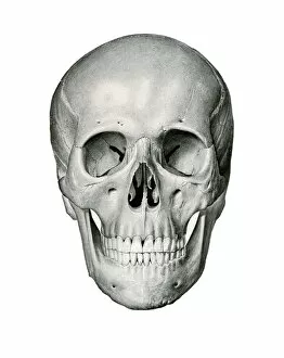
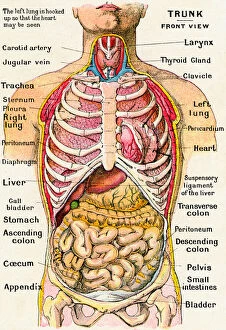
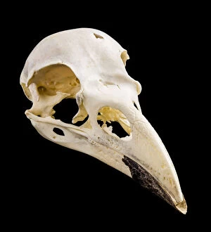
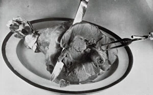
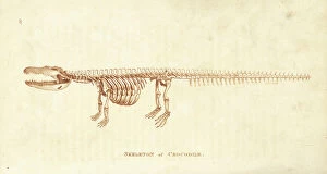
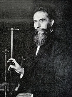
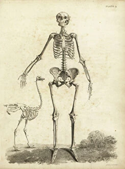
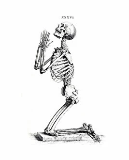
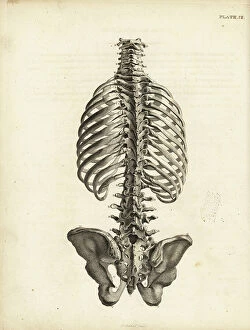
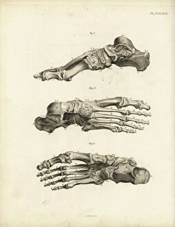
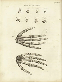
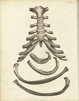
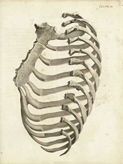
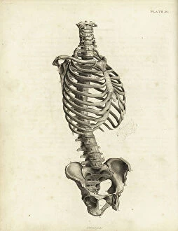
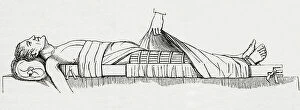
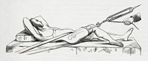
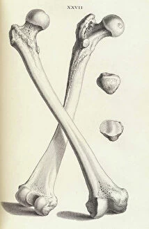
"Bones: Unveiling the Hidden Structures of Life" Delving into the depths of human anatomy, Leonardo da Vinci's "Skull Anatomy" showcases the intricate beauty and complexity of bones. A graceful greyhound's skeleton reveals the elegance and agility that lies beneath its sleek exterior. Behind-the-scenes in Bristol, Christopher Ryan transforms into Mike from "The Young Ones, " a character whose humor is as sharp as his bone structure. Captured on an X-ray, a broken wrist bone serves as a reminder of our vulnerability and resilience in the face of injury. The majestic skull of a horse stands as a testament to their strength and power, reminding us of their historical significance alongside humans. Even skeletons enjoy some rough play. Witnessing a rugby-playing skeleton brings forth both amusement and admiration for these resilient structures. Knapped Flint Tools take us back in time, showcasing how early humans utilized bones to shape tools for survival and progress. Peering through panoramic dental X-rays unravels secrets hidden within our mouths – teeth anchored by strong bones supporting our smiles. Our bodies are marvels intertwined with arteries that nourish every cell while sturdy bones provide support - truly works of art designed by nature itself. In an act of kindness, Chef Andrew Schillar shares his culinary delight with man's best friend - offering them not just food but also joyous moments gnawing on a bone outside his kitchen sanctuary. Illustrated through wood engraving in 1851, "Death as Assassin" reminds us that even though life may be fleeting, it is ultimately connected to the skeletal framework we all share. Modern technology unveils our innermost secrets with full-body scans like MRI scans – exposing not only flesh but also revealing the silent symphony orchestrated by our interconnected bones.