
Bronchial Tree Collection
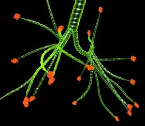
The bronchial tree is a fascinating part of the human respiratory system, consisting of the bronchus and bronchial tubes


For sale as Licensed Images
Choose your image, Select your licence and Download the media
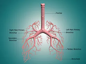
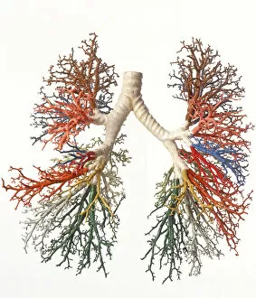
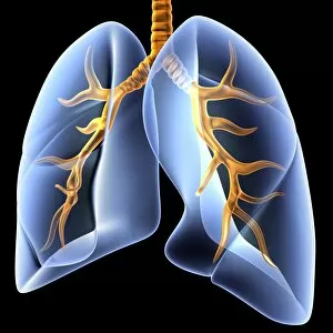
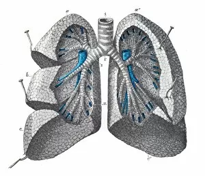
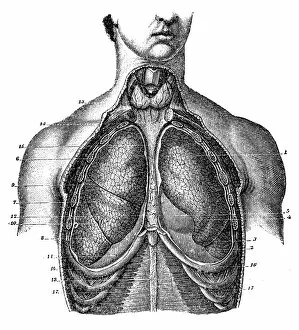
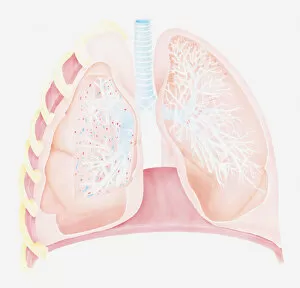
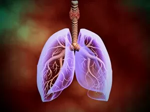
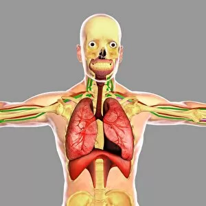
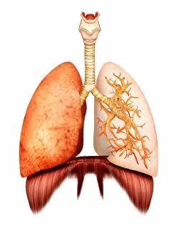
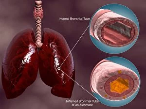
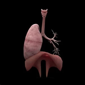
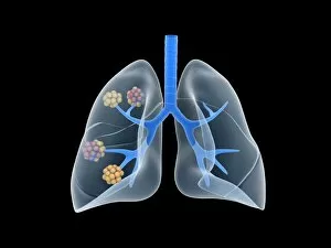
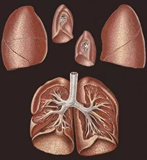
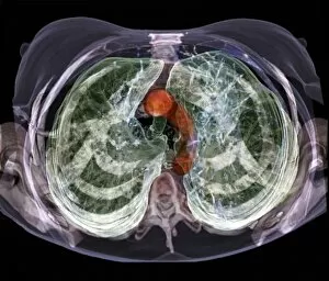
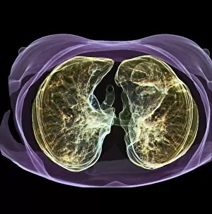
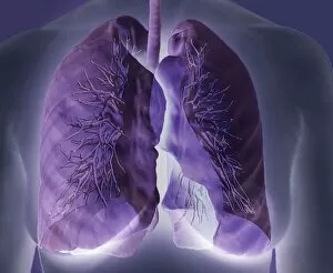
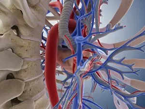
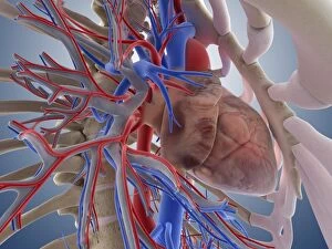
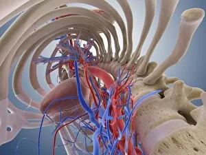
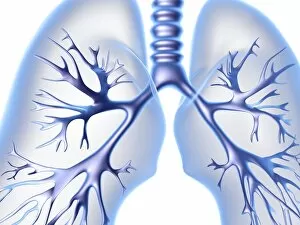
The bronchial tree is a fascinating part of the human respiratory system, consisting of the bronchus and bronchial tubes. It resembles a complex network of branches that extend deep into our lungs, allowing us to breathe in life-giving oxygen. If you were to examine a model of the bronchial tree, you would be amazed at its intricate design. Each branch leads to smaller and smaller tubes, eventually culminating in tiny structures called lung alveoli. These alveoli resemble a delicate tree with countless leaves where gas exchange takes place. Looking at an engraving from 1899 depicting pulmonary mass, we can appreciate how scientists have long been captivated by the intricacies of this vital organ. The illustration showcases the complexity and beauty hidden within our lungs. In cross-sectional biomedical illustrations of human lungs, we gain insight into their internal structure. We see how air passes through the trachea and enters the lungs through branching pathways known as bronchioles. This detailed view allows us to understand both normal lung function and abnormalities like asthma. One striking image shows transparency in an illustration featuring the trachea with lungs. This unique perspective offers a glimpse into how air flows through this crucial pathway during respiration. Studying these images reminds us just how remarkable our respiratory system truly is. From front views showcasing its anatomy to cross-sections revealing its inner workings, it becomes clear that every breath we take relies on this intricate network within our bodies. So next time you take a deep breath, remember all those branches forming your own personal bronchial tree – silently working together to keep you alive and thriving.