Bronchiole Collection (#2)

The bronchiole is a vital component of the respiratory system, specifically within the anatomy of the bronchus and bronchial tubes

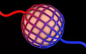
Background imageBronchiole Collection: Conceptual image of alveolus

Conceptual image of alveolus

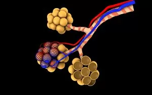
Background imageBronchiole Collection: Conceptual image of alveoli

Conceptual image of alveoli

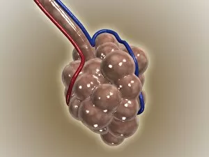
Background imageBronchiole Collection: Close-up of alveolus

Close-up of alveolus
The alveoli are the final branchings of the respiratory tree and act as the primary gas exchange units of the lung

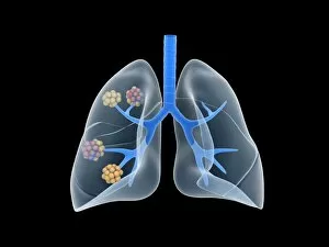
Background imageBronchiole Collection: Conceptual image of human lungs

Conceptual image of human lungs

Background imageBronchiole Collection: Three dimensional view of the male respiratory system

Three dimensional view of the male respiratory system, close-up

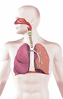
Background imageBronchiole Collection: Cross section of human respiratory system

Cross section of human respiratory system

Background imageBronchiole Collection: Illustration of respiratory system

Illustration of respiratory system
Medicine: Human anatomy, respiratory system. Drawing

Background imageBronchiole Collection: Lung, X-ray F008 / 3219

Lung, X-ray F008 / 3219
Lung. Coloured X-ray showing the blood vessels in a lung

Background imageBronchiole Collection: Human lungs, artwork F008 / 3305

Human lungs, artwork F008 / 3305
Human lungs, computer artwork

Background imageBronchiole Collection: Human lungs, artwork F008 / 3304

Human lungs, artwork F008 / 3304
Human lungs, computer artwork

Background imageBronchiole Collection: Lung, X-ray F008 / 3220

Lung, X-ray F008 / 3220
Lung. Coloured X-ray showing the blood vessels in a lung

Background imageBronchiole Collection: Lungs anatomy, artwork

Lungs anatomy, artwork
Lungs anatomy, computer artwork. At top left is the trachea (windpipe, white), which splits into two bronchi, one for each lung (blue)

Background imageBronchiole Collection: Respiratory system, artwork C015 / 6453

Respiratory system, artwork C015 / 6453
Respiratory system. Computer artwork showing the organs of the human respiratory system. At top is the larynx (voicebox), which leads onto the trachea (windpipe, white tube vertical)

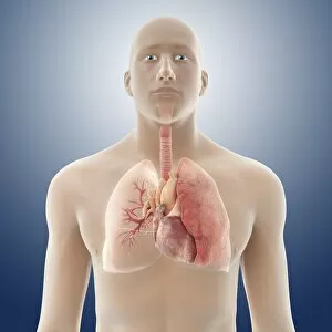
Background imageBronchiole Collection: Heart and lung anatomy, artwork C016 / 2909

Heart and lung anatomy, artwork C016 / 2909
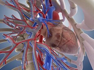
Heart and lung anatomy. Artwork of an anterior (frontal) view of the heart and lungs in a male body. One lung is shown at right. The bronchioles (airways) inside the lungs are shown at left

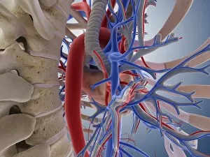
Background imageBronchiole Collection: Heart and lung anatomy, artwork C016 / 2904

Heart and lung anatomy, artwork C016 / 2904
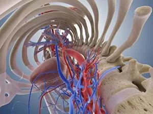
Heart and lung anatomy. Artwork of a posterior (from behind) view of the heart and lungs. The heart (mostly obscured) is a hollow muscle that pumps blood around the body and to the lungs

Background imageBronchiole Collection: Heart-lung system, artwork C013 / 4412

Heart-lung system, artwork C013 / 4412
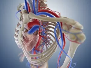
Heart-lung system, computer artwork. The heart (centre) is a hollow muscle that pumps blood around the body. Arteries, which carry oxygenated blood around the body, are red

Background imageBronchiole Collection: Heart-lung system, artwork C013 / 4411

Heart-lung system, artwork C013 / 4411
Heart-lung system, computer artwork. The heart (centre) is a hollow muscle that pumps blood around the body. Arteries, which carry oxygenated blood around the body, are red

Background imageBronchiole Collection: Heart-lung system, artwork C013 / 4410

Heart-lung system, artwork C013 / 4410
Heart-lung system, computer artwork. The heart (lower centre) is a hollow muscle that pumps blood around the body. Arteries, which carry oxygenated blood around the body, are red

Background imageBronchiole Collection: Heart-lung system, artwork C013 / 4408

Heart-lung system, artwork C013 / 4408
Heart-lung system, computer artwork. The heart (centre) is a hollow muscle that pumps blood around the body. Arteries, which carry oxygenated blood around the body, are red

Background imageBronchiole Collection: Heart-lung system, artwork C013 / 4409

Heart-lung system, artwork C013 / 4409
Heart-lung system, computer artwork. The heart (centre left) is a hollow muscle that pumps blood around the body. Arteries, which carry oxygenated blood around the body, are red

Background imageBronchiole Collection: Heart-lung system, artwork C013 / 4407

Heart-lung system, artwork C013 / 4407
Heart-lung system, computer artwork. The heart (centre left) is a hollow muscle that pumps blood around the body. Arteries, which carry oxygenated blood around the body, are red

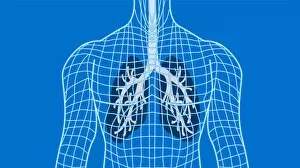
Background imageBronchiole Collection: Lungs

Lungs in a wireframe body, computer artwork. The windpipe (trachea, grey) runs downwards from the neck. It branches (upper centre) into two bronchi, one for each of the two lungs

Background imageBronchiole Collection: SEM of lung alveoli

SEM of lung alveoli
Lung alveoli. Coloured Scanning Electron Micro- graph (SEM) of lung tissue, showing alveoli and a pulmonary blood vessel. At centre is the lumen of a large blood vessel (yellow)

Background imageBronchiole Collection: Bronchioles

Bronchioles
Bronchiole network, composite image. The trachea (windpipe, not seen) branches into two bronchi, which enter the pair of lungs

Background imageBronchiole Collection: Lung, X-ray

Lung, X-ray
Lung. Coloured bronchography (X-ray) of a healthy human lung. A contrast medium has been added to show the network of airways (green) in the right lung

Background imageBronchiole Collection: Lung bronchogram, coloured X-ray

Lung bronchogram, coloured X-ray
Healthy lung. Coloured bronchogram x-ray of a side (lateral) view of a healthy human lung. The vertebrae of the spinal column are seen at left

Background imageBronchiole Collection: Lungs, CT scans

Lungs, CT scans
Lungs. Sequence of six coloured axial CT (computed tomography) scans through the upper chest of a 52- year-old woman. The front of the chest is at top, and the right of the body is at left

Background imageBronchiole Collection: Computer artwork of healthy human lungs

Computer artwork of healthy human lungs

Background imageBronchiole Collection: Lung airways

Lung airways. Computer-enhanced image of a resin cast of the airways in the lungs. The trachea (windpipe, top centre) divides into two main bronchi

Background imageBronchiole Collection: Emphysema of the lungs, CT scan

Emphysema of the lungs, CT scan
Emphysema. Coloured 3-D computed tomography (CT) scan of the lungs, showing advanced emphysema of the right lung (left). Emphysema is a disease in which the tiny lung air sacs (alveoli)

Background imageBronchiole Collection: Artwork of asthmatic respiratory system on inhaler

Artwork of asthmatic respiratory system on inhaler
Asthma. Artwork of part of the respiratory system, seen on an aerosol inhaler (blue), showing the effects of asthma. The bronchi, covered in bands of cartilage (pink)

Background imageBronchiole Collection: Asthma, artwork

Asthma, artwork
Asthma. Computer artwork of allergens (green) in the airways of the lungs. Allergens, such as dust and pollen, can trigger an asthma attack

Background imageBronchiole Collection: Childhood asthma

Childhood asthma. Artwork of a childs respiratory system showing the mucus (yellow) and narrowing of the airways (bronchioles) in paediatric asthma

Background imageBronchiole Collection: Artwork of bronchodilator action in asthma

Artwork of bronchodilator action in asthma
Asthma treatment. Artwork showing the action of a bronchodilator drug breathed into the respiratory system from an aerosol inhaler to treat asthma

Background imageBronchiole Collection: Bronchitis, X-ray

Bronchitis, X-ray
Bronchitis. Coloured bronchography (X-ray) of swollen bronchioles (orange) in the right lung. Bronchitis causes the network of airways in the lungs to become swollen and produce mucus

Background imageBronchiole Collection: Asthma

Asthma. Computer artwork of allergens (blue and green) amongst alveoli (tiny air sacs, beige) in a lung. Allergens, such as dust and pollen, can trigger an asthma attack

Background imageBronchiole Collection: Artwork of the respiratory system of an asthmatic

Artwork of the respiratory system of an asthmatic
Asthma. Cut-away illustration of the respiratory system showing the effects of asthma. The trachea (windpipe) is at top and the alveoli, air sacs (white), are at bottom

Background imageBronchiole Collection: Artwork of bronchodilator drug in bronchiole

Artwork of bronchodilator drug in bronchiole

Background imageBronchiole Collection: Artwork of inflamed bronchial epithelium in asthma

Artwork of inflamed bronchial epithelium in asthma
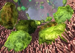
Bronchial inflammation in asthma. Artwork of bron- chial epithelial tissue inflamed by asthma. A thick layer of mucus (green) covers the tissue

Background imageBronchiole Collection: Alveoli in the lung, artwork

Alveoli in the lung, artwork
Alveoli in the lung. Computer artwork showing the alveoli (spheres) and bronchioles (tubes) of the lungs. The alveoli are the tiny air sacs of the lungs where gases are exchanged between the air in

Background imageBronchiole Collection: Pulmonary lobule, artwork

Pulmonary lobule, artwork
Pulmonary lobule. Computer artwork of a section through an area of lung tissue. A pulmonary lobule is a unit of lung tissue that includes a bronchiole, alveolar ducts, sacs, and alveoli

Background imageBronchiole Collection: Heart and lungs, historical illustration

Heart and lungs, historical illustration. 19th Century hand coloured lithographic print showing the lungs (left and right) situated either side of the heart (lower centre)

Background imageBronchiole Collection: Respiratory anatomy, 19th Century artwork

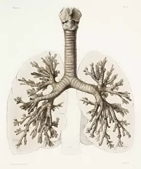
Respiratory anatomy, 19th Century artwork. Historical hand coloured lithographic print showing the trachea (wind pipe, vertical) running down from the larynx (voicebox)

Background imageBronchiole Collection: Respiratory anatomy, artwork

Respiratory anatomy, artwork
Respiratory anatomy. Computer artwork showing the structure of the human respiratory system, including the lungs (left and right), larynx (voicebox, upper centre) and trachea (windpipe, purple)

Background imageBronchiole Collection: Respiratory tract, artwork

Respiratory tract, artwork
Respiratory tract. Computer artwork showing the various stages and structures of the human respiratory tract. Air is drawn in through the oral and/or nasal cavities, and passes down the trachea

Background imageBronchiole Collection: Lung alveolus, artwork

Lung alveolus, artwork
Lung alveolus, computer artwork. Alveoli are the site of gas exchange in the lungs of mammals, where oxygen is taken up and carbon dioxide released

Background imageBronchiole Collection: Human lungs

Human lungs. Anatomical artwork of the human lungs and respiratory system. The heart (which has been removed here) is located between the lungs

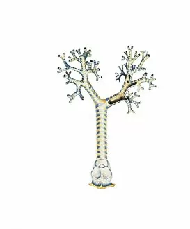
Background imageBronchiole Collection: Bronchus and bronchioles, artwork

Bronchus and bronchioles, artwork
Bronchus and bronchioles. Artwork of the anatomical structure of the bronchi and their bronchioles, the main aspects of the respiratory system


For sale as Licensed Images

Choose your image, Select your licence and Download the media

The bronchiole is a vital component of the respiratory system, specifically within the anatomy of the bronchus and bronchial tubes. These delicate structures are responsible for carrying air into and out of the lungs, ensuring proper oxygenation and removal of carbon dioxide. When examining an X-ray of the lung, one can observe these intricate passageways branching off from the main bronchi. They resemble a tree-like structure, with smaller branches known as bronchioles extending deeper into the lung tissue. In understanding male chest anatomy, it becomes evident that the they are nestled within the thorax alongside other essential organs such as the heart, veins, arteries, and lungs. This interconnectedness highlights their crucial role in maintaining respiratory function. Examining internal organ anatomy reveals how intricately woven together systems like respiration and circulation truly are. The human circulatory system relies on efficient oxygen exchange facilitated by healthy bronchioles to ensure optimal functioning throughout all bodily processes. A cutaway diagram provides a comprehensive view of how our respiratory system operates. It showcases how each individual lung is connected to its respective primary bronchus through numerous secondary and tertiary branches called bronchioles. Conceptual images further emphasize this interplay between different systems within our bodies. They illustrate how internal organs belonging to both respiratory and digestive systems coexist harmoniously while performing their distinct functions. Studying pulmonary embolism sheds light on potential complications that can arise within these pathways. An embolus traveling through veins may reach its final destination in either heart or lungs via various routes involving intricate networks of arteries and veins surrounding them. Understanding arterial-venous relationships across our body aids in comprehending blood flow dynamics associated with respiration. Arteries carry freshly oxygenated blood from lungs to tissues while veins return deoxygenated blood back for reoxygenation – all made possible by well-functioning bronchioles among other components involved in gas exchange.