
Bronchus Collection (#2)
The bronchus, a vital component of the human respiratory system, plays a crucial role in delivering oxygen to our lungs


For sale as Licensed Images
Choose your image, Select your licence and Download the media
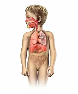
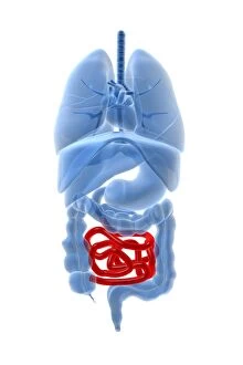
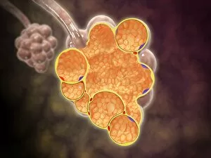
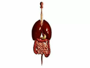
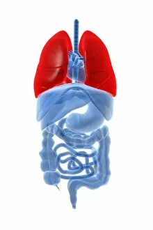
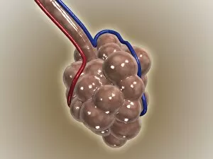
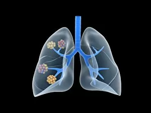
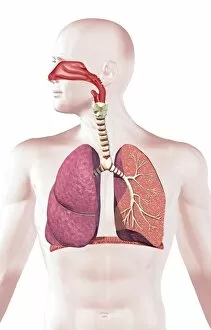
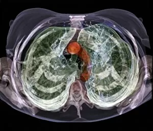
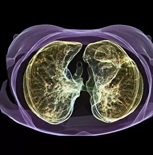
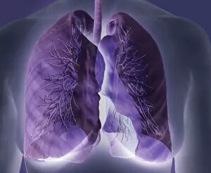
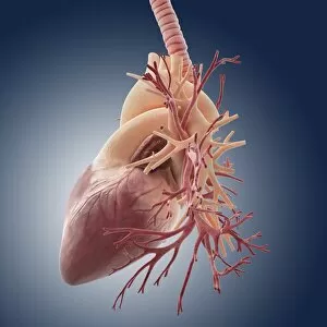
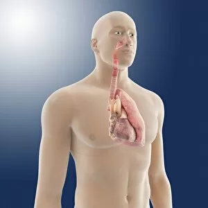
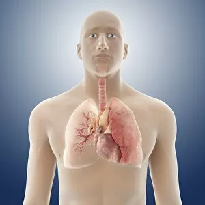
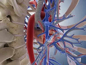
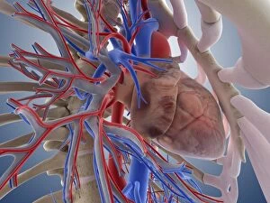
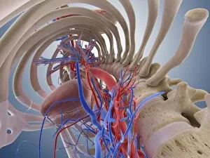
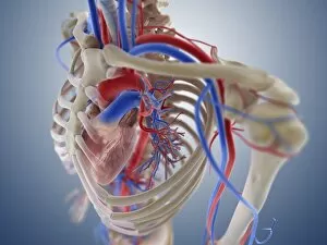
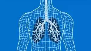
The bronchus, a vital component of the human respiratory system, plays a crucial role in delivering oxygen to our lungs. This intricate network of airways can be visualized through various illustrations and diagrams. In a lung diagram, the bronchus is depicted alongside other respiratory structures such as the oral cavity, nasal cavity, larynx, trachea, and lungs. This illustration highlights how these interconnected parts work together seamlessly to facilitate breathing. When examining the anatomy of the bronchus and bronchial tubes, one can appreciate their complex structure and function. These slender passageways branch out like tree branches within our lungs, ensuring that every nook and cranny receives life-giving oxygen. However, certain conditions can affect the health of our bronchi. Cystic fibrosis is one such ailment that impacts these air passages by causing them to become thickened and clogged with mucus. Understanding this disease helps us comprehend why individuals with cystic fibrosis may experience difficulty breathing. An X-ray image showcasing human lungs allows us to visualize not only their overall shape but also provides insight into how the bronchi traverse throughout this essential organ. The intricacies captured in this image remind us of just how remarkable our internal systems truly are. Exploring male or female body anatomies further reveals how the bronchi intertwine with other organs within our chests. In males specifically, an illustration demonstrates thorax anatomy featuring heart veins arteries along with prominent lungs - all connected by branching bronchi. Additionally, understanding blood supply becomes crucial when studying both cardiovascular health and respiratory systems simultaneously. The Heart and Bronchial Arteries facsimile from Windsor book showcases pen-and-ink artistry while highlighting these important connections between circulation and respiration. Ultimately though it's important for everyone to grasp basic knowledge about human lung anatomy as it directly relates to everyday living; after all healthy functioning lungs are fundamental for survival.