
Calcaneus Collection
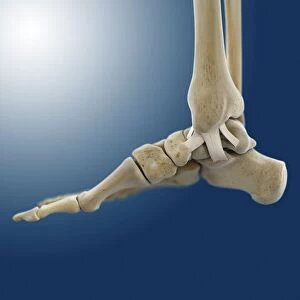
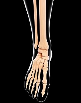
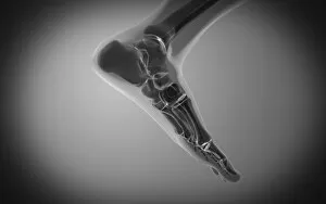
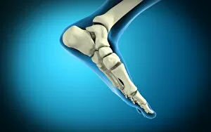
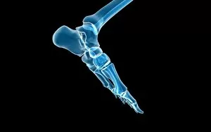
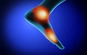
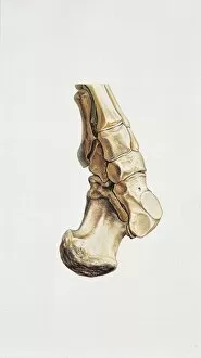
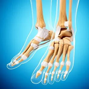
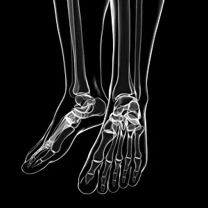
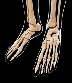
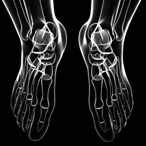
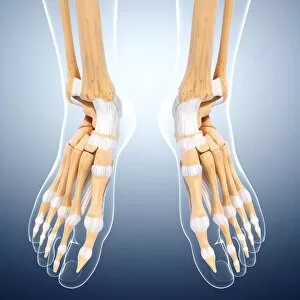
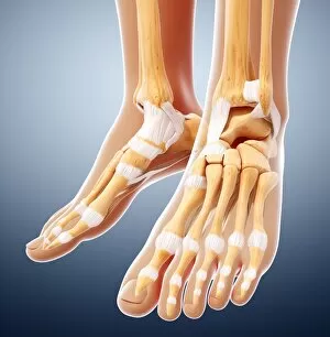
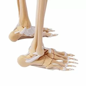
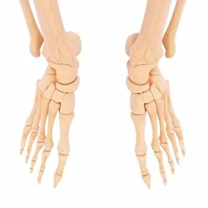
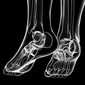
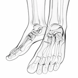
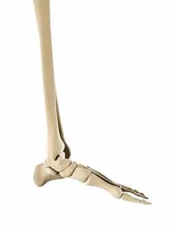
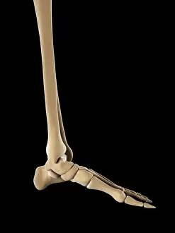
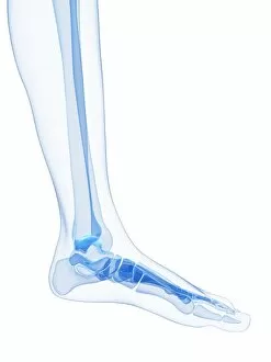
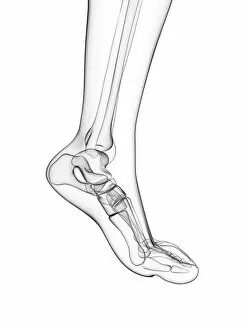
The calcaneus, also known as the heel bone, is a crucial part of the human foot anatomy. Located at the back of the foot, it forms the foundation for our every step

For sale as Licensed Images
Choose your image, Select your licence and Download the media
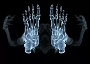
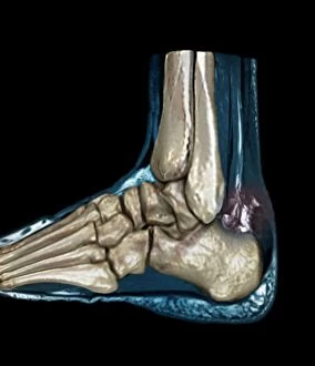
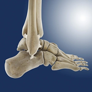
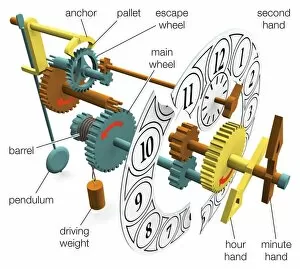
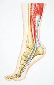
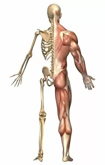
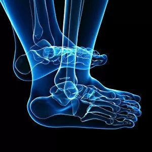
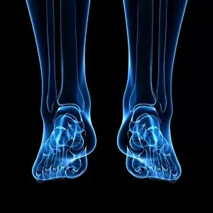
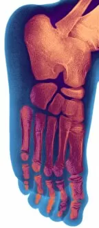
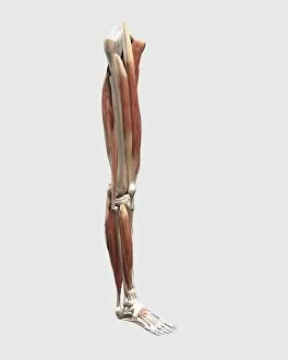
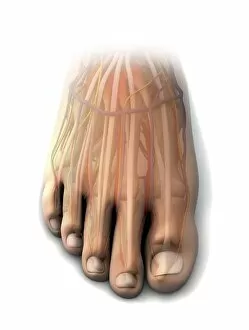
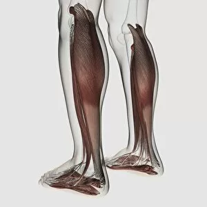
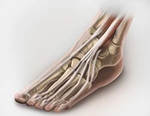
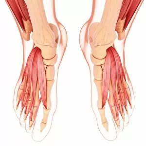
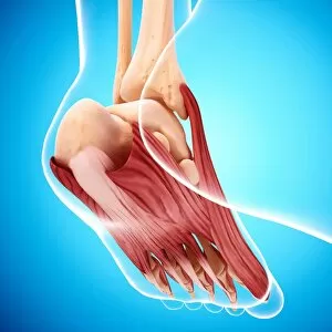
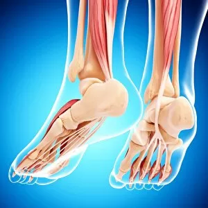
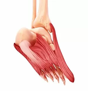
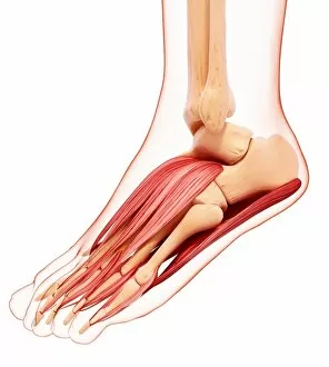
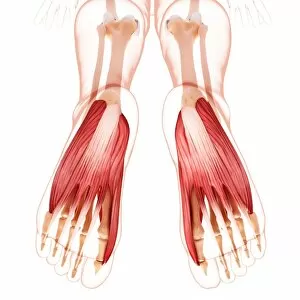
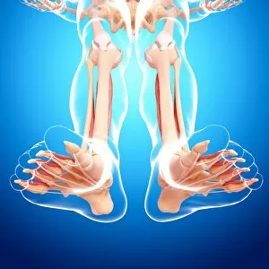
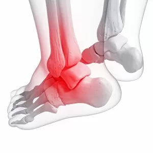
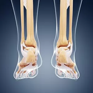
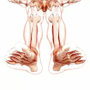
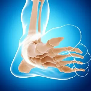
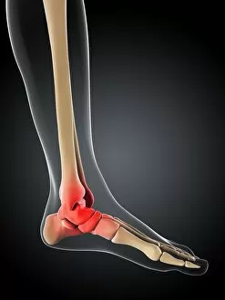
The calcaneus, also known as the heel bone, is a crucial part of the human foot anatomy. Located at the back of the foot, it forms the foundation for our every step. In an X-ray artwork showcasing a skeleton from below, we can see this sturdy bone in all its glory. But there's more to the calcaneus than meets the eye. When we delve deeper into its intricate structure, an MRI image reveals a ruptured Achilles tendon nearby. This injury serves as a reminder of how important it is to take care of our feet and avoid overexertion. Artwork depicting outer ankle ligaments showcases their role in providing stability and preventing sprains. Similarly, inner ankle ligaments are highlighted in another artwork, emphasizing their significance in maintaining balance during movement. Just like a pendulum clock swings with precision, our feet flex effortlessly thanks to well-coordinated bones and relevant muscles revealed when flexed. It's fascinating how these components work together harmoniously to enable us to walk or run smoothly. In front view images of both skeletal and muscular systems, we gain insight into how leg bones support body weight while muscles provide strength and flexibility. The complexity of this system becomes evident as we observe various artworks illustrating different aspects such as leg bones or foot bones individually. Even children's feet have been studied through X-rays, reminding us that understanding foot development from an early age is essential for proper growth and prevention of future issues.