
Cancer Collection (#9)
"Cancer: A Journey Through Time and Cells" Step back in time to ancient Egypt with a captivating map showcasing the rich history of this enigmatic civilization


For sale as Licensed Images
Choose your image, Select your licence and Download the media
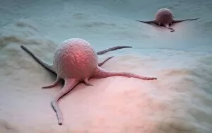
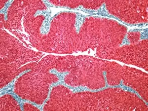
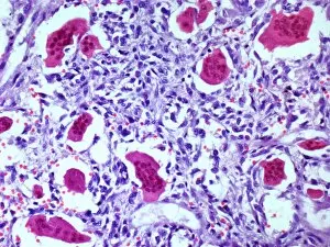
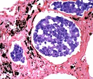
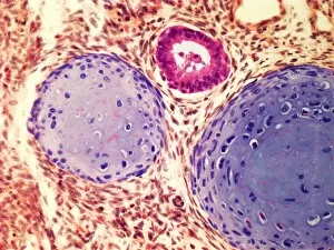
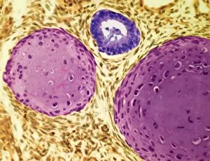
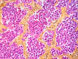
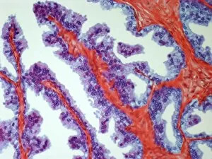
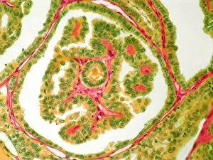
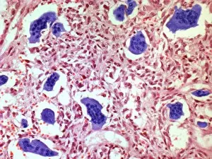
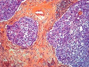
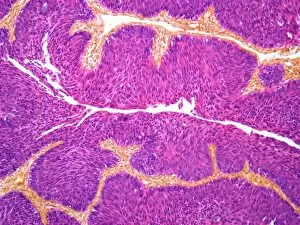
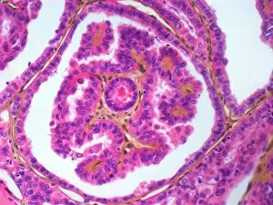
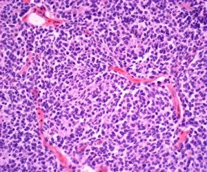
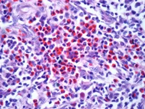
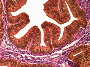
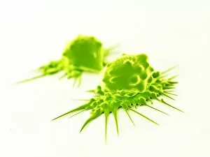
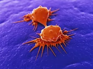
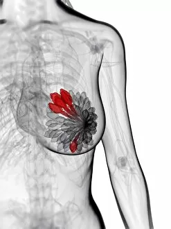
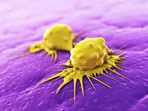
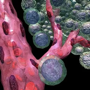
"Cancer: A Journey Through Time and Cells" Step back in time to ancient Egypt with a captivating map showcasing the rich history of this enigmatic civilization. Amidst the grandeur, let us delve into the intricate world of cancer. Witness the battle within our bodies as T lymphocytes fiercely combat cancer cells under an SEM microscope. This microscopic view reveals the relentless fight that takes place daily, reminding us of both resilience and vulnerability. Travel across continents to California's vibrant city, San Diego, where the iconic US Midway stands tall as a museum and event venue. Just like this majestic ship, we navigate through life's challenges with strength and determination when faced with cancer. In London, marvel at the mesmerizing sight of the illuminated pink London Eye against the backdrop of River Thames. It symbolizes hope and unity in raising awareness for breast cancer – a reminder that together we can conquer any obstacle. From ancient artistry to modern interpretations, Eugene Samuel Grasset's masterpiece "July: Harvesting and Sheep Shearing" transports us to simpler times when nature dictated our lives. Cancer reminds us that life is precious; it urges us to cherish every moment spent amidst bountiful fields or beneath clear blue skies. Centuries ago, frescoes depicted June as a month filled with abundance and joyous celebrations. Today we honor those affected by cancer during this month – celebrating their strength while striving for breakthroughs in research and treatment options. Peer into medical science through micrographs capturing acute promyelocytic leukemia or ovarian cancer cells under light microscopy. These images serve as reminders that behind every diagnosis lies immense courage from patients battling these diseases head-on. Just like diverse crab varieties found along coastlines worldwide, each person's experience with cancer is unique yet interconnected by shared struggles and triumphs. Let empathy guide our understanding as we support one another on this challenging journey. Finally, explore your zodiac horoscope star sign – an emblem of individuality and cosmic influence.