
Cardiovascular Collection

"Exploring the Intricacies of the Cardiovascular System

Background imageCardiovascular Collection: Diagram of the circulation of blood as understood after William Harvey's work

Diagram of the circulation of blood as understood after William Harvey's work
5309470 Diagram of the circulation of blood as understood after William Harvey's work, showing the Lesser Circulation (Pulmonary Circulation)

Background imageCardiovascular Collection: Engraving depicting a human heart, 18th century

Engraving depicting a human heart, 18th century
5309305 Engraving depicting a human heart, 18th century; (add.info.: Engraving depicting a human heart. Dated 18th century 01/02/1764); Universal History Archive/UIG

Background imageCardiovascular Collection: Coronary arteries, illustration

Coronary arteries, illustration
Illustration of coronary arteries of the heart

Background imageCardiovascular Collection: Female heart anatomy, illustration

Female heart anatomy, illustration
Female heart anatomy, computer illustration

Background imageCardiovascular Collection: Posterior view of a normal heart and its arteries

Posterior view of a normal heart and its arteries
Detail, Anatomy, Diagram, Muscle, Heart, Illustration, Medical, Biology, Cardiac, Rear View, Cardiovascular, Cut Out, White Background, Vertical, Artwork, Color Image, Close Up

Background imageCardiovascular Collection: Human cardiovascular system

Human cardiovascular system, illustration

Background imageCardiovascular Collection: Babys heart and circulatory system, illustration

Babys heart and circulatory system, illustration
Illustration of a babys heart and circulatory system

Background imageCardiovascular Collection: Front view of a normal heart and its cornonary arteries and veins

Front view of a normal heart and its cornonary arteries and veins
Detail, Anatomy, Diagram, Heart, Illustration, Medical, Vein, Biology, Cardiac, Veins, Cardiovascular, Cut Out, White Background, Vertical, Artwork, Color Image, Front View, Close Up

Background imageCardiovascular Collection: Female anatomy

Female anatomy
http://csaimages.com/images/istockprofile/csa_vector_dsp.jpg

Background imageCardiovascular Collection: Veins and arteries in the head, plate from Recherches Anatomiques, Physiologiques

Veins and arteries in the head, plate from Recherches Anatomiques, Physiologiques
CHT235023 Veins and arteries in the head, plate from Recherches Anatomiques, Physiologiques, et Pathologiques sur le System Veineux by Gilbert Breschet (1784-1845) engraved by Engelmann, Paris

Background imageCardiovascular Collection: Internal organs of a woman, plate from Recherches Anatomiques, Physiologiques

Internal organs of a woman, plate from Recherches Anatomiques, Physiologiques
CHT235024 Internal organs of a woman, plate from Recherches Anatomiques, Physiologiques, et Pathologiques sur le System Veineux by Gilbert Breschet (1784-1845) engraved by Engelmann, Paris

Background imageCardiovascular Collection: Heart with coronary vessels, illustration

Heart with coronary vessels, illustration
Heart with coronary blood vessels, computer illustration

Background imageCardiovascular Collection: Heart bypass graft, artwork

Heart bypass graft, artwork
Heart bypass graft. Computer artwork of a heart that has had a blockage of the coronary arteries treated by coronary artery bypass graft (CABG) surgery

Background imageCardiovascular Collection: Human heart, illustration

Human heart, illustration
Human heart, computer illustration

Background imageCardiovascular Collection: Male heart, illustration

Male heart, illustration
Illustration of male heart and arteries

Background imageCardiovascular Collection: Female vascular system, illustration

Female vascular system, illustration
Female vascular system, computer illustration

Background imageCardiovascular Collection: Cross section of a normal heart

Cross section of a normal heart
Detail, Anatomy, Diagram, Muscle, Heart, Illustration, Biology, Chamber, Cardiac, Atrium, Cardiovascular, Cut Out, White Background, Vertical, Artwork, Cross Section, Color Image, Close Up

Background imageCardiovascular Collection: Cross section of a normal heart

Cross section of a normal heart
Detail, Anatomy, Diagram, Muscle, Heart, Illustration, Medical, Biology, Atrium, Cardiovascular, Cut Out, White Background, Vertical, Artwork, Cross Section, Color Image, Close Up

Background imageCardiovascular Collection: Resting T lymphocytes

Resting T lymphocytes. Coloured scanning electron micrograph (SEM) of resting T lymphocytes from a human blood sample. T lymphocytes, or T cells

Background imageCardiovascular Collection: Grays Anatomy - carotid artery

Grays Anatomy - carotid artery
Two pages from the second edition of Grays Anatomy dealing with the common carotid artery. With hand-coloured illustration showing the blood vessels of the neck

Background imageCardiovascular Collection: Female anatomy, historical model

Female anatomy, historical model
Female anatomy. 1920s anatomical model showing the internal organs of the female upper body with the intestines, stomach and liver removed

Background imageCardiovascular Collection: Cardiovascular system Leonardo da vincis drawing

Cardiovascular system Leonardo da vincis drawing
Cardiovascular systems drawing. In 1513 Leonardo da Vinci to study the heart and the circulatory system through animal dissections. Windsor, Royal Library 19073, 1510-1513

Background imageCardiovascular Collection: The veins of the human body

The veins of the human body
A woodcut showing the veins of the human body which are just under the skin. From: Mikrokosmographia: a description of the body of man, together with the controversies and figures thereto belonging

Background imageCardiovascular Collection: Neck and head arteries, MRA scan C016 / 6449

Neck and head arteries, MRA scan C016 / 6449
Neck and head arteries. Coloured magnetic resonance angiography (MRA) scan of a normal carotid system of a 36 year old patient

Background imageCardiovascular Collection: CARREL, Alexis (1873 - 1944). French surgeon

CARREL, Alexis (1873 - 1944). French surgeon and biologist. Nobel Prize in Medicine in 1912. Oil

Background imageCardiovascular Collection: The nerves of the arm in Leprosy

The nerves of the arm in Leprosy
Plate IX: A depiction of the nerves of the arm in Leprosy. From: On leprosy and elephantiasis. Date: 1874

Background imageCardiovascular Collection: Two anatomical figures, one male and the other female

Two anatomical figures, one male and the other female
Title page of Mikrokosmographia with two anatomical figures, one male and the other female. Fom: Mikrokosmographia: a description of the body of man

Background imageCardiovascular Collection: Marey type sphygmograph, 19th century C017 / 6933

Marey type sphygmograph, 19th century C017 / 6933
Marey type sphygmograph, made in France around 1870. This instrument is boxed, made from gilt metal and has a clockwork mechanism

Background imageCardiovascular Collection: Boulitte oscillometer, circa 1910 C017 / 6940

Boulitte oscillometer, circa 1910 C017 / 6940
Boulitte oscillometer, dating from 1910, France. This device is based on an invention by French physiologist, Michel Victor Pachon (1867-1938)

Background imageCardiovascular Collection: Laennec monaural stethoscope, circa 1820 C017 / 6951

Laennec monaural stethoscope, circa 1820 C017 / 6951
English monaural stethoscope, made out of fruitwood, dating from the year 1820. French physician, Rene Theophile Hyacinthe Laennec invented the monaural stethoscope in 1816

Background imageCardiovascular Collection: Monaural stethoscopes, 19th century C017 / 6931

Monaural stethoscopes, 19th century C017 / 6931
Five monaural stethoscopes dating from between 1860 and 1890. These are made from ebony and fruitwood. French physician, Rene Theophile Hyacinthe Laennec invented the monaural stethoscope in 1816

Background imageCardiovascular Collection: Phonendoscope, circa 1900 C017 / 6955

Phonendoscope, circa 1900 C017 / 6955
Phonendoscope dating from around 1900, England. This device was used in the early 20th century for listening to internal sounds of the body (auscultation)

Background imageCardiovascular Collection: Extra long monaural stethoscope, circa 18 C017 / 6948

Extra long monaural stethoscope, circa 18 C017 / 6948
Unusual extra long monaural stethoscope dating from 1860. This piece was made in England out of fruitwood. French physician

Background imageCardiovascular Collection: Sphygmometer by Collin, circa 1900 C017 / 6972

Sphygmometer by Collin, circa 1900 C017 / 6972
Sphygmometer in its case. This sphygmometer was made in France, around 1900 by Collin, a well known French instrument maker

Background imageCardiovascular Collection: Drainage trocar with cannulae, circa 1860 C017 / 6949

Drainage trocar with cannulae, circa 1860 C017 / 6949
Dr. Southeys drainage trocar with 3 cannulae in their case. This set dates from 1860 and is made by Arnold and Sons, England

Background imageCardiovascular Collection: Sphygmometer by Collin, circa 1900 C017 / 6966

Sphygmometer by Collin, circa 1900 C017 / 6966
Sphygmometer made in France, around 1900 by Collin, a well known French instrument maker. Sphygmometers such as this one were used to demonstrate arterial blood pressure using an aneroid manometer

Background imageCardiovascular Collection: Richardsons sphygmograph, circa 1882

Richardsons sphygmograph, circa 1882
Richardsons sphygmograph, in its case, dating from around 1882. This sphygmograph is nickel and was made in England by Allen and Hanbury

Background imageCardiovascular Collection: Modified Dudgeon sphygmograph, circa 1890 C017 / 6943

Modified Dudgeon sphygmograph, circa 1890 C017 / 6943
Modification of Dudgeon sphygmograph with tambour dating from around 1890, England. This instrument is made of nickel and has a clockwork mechanism

Background imageCardiovascular Collection: Sphygmomanometer, circa 1920 C017 / 6945

Sphygmomanometer, circa 1920 C017 / 6945
Martins Improved Sphygmomanometer in its case, dating from 1920. This instrument is made in glass and mercury by S. Maw, Son and Sons


For sale as Licensed Images

Choose your image, Select your licence and Download the media

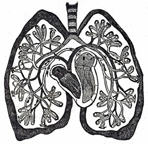
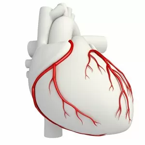
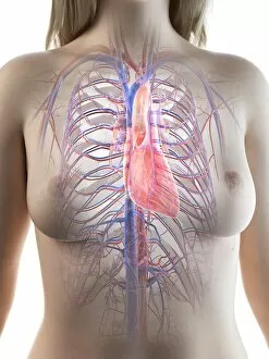
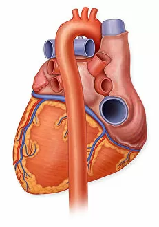
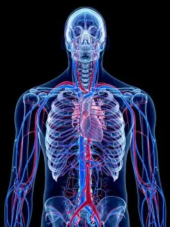
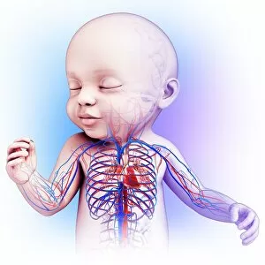
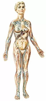
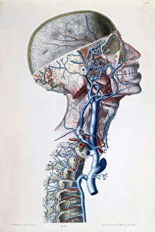
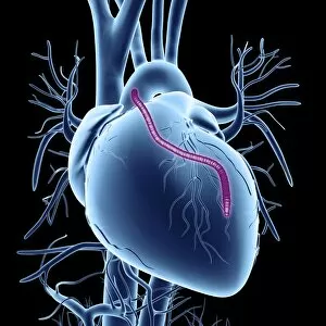
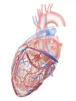
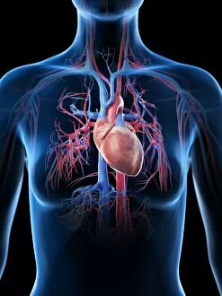
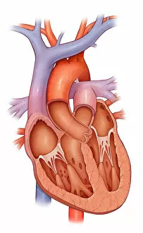
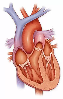
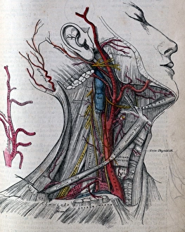
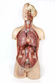
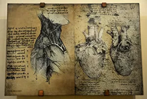
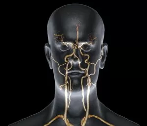
"Exploring the Intricacies of the Cardiovascular System: A Journey through Anatomy and Physiology" Delve into the intricate network of coronary arteries with a detailed illustration, unraveling their vital role in nourishing the heart muscle. Discover the wonders of female heart anatomy through an exquisite illustration, showcasing its unique structure and function. Take a posterior view of a normal heart and its arteries, marveling at this remarkable organ's ability to pump life-giving blood throughout our bodies. Dive into Grays Anatomy as we explore the carotid artery, one of the major vessels supplying oxygen-rich blood to our brain – truly a lifeline for human cognition. Embark on an enlightening journey through the human cardiovascular system, uncovering its complexity and understanding how it sustains our very existence. Witness the incredible development of a baby's heart and circulatory system through an enchanting illustration that highlights nature's miraculous design. Gain insight into female anatomy as we examine internal organs intricately depicted in stunning detail, shedding light on their interconnectedness within our cardiovascular framework. Unravel the mesmerizing web of veins coursing throughout our bodies with an awe-inspiring visual representation that showcases their crucial role in returning deoxygenated blood back to the heart. Peer into Recherches Anatomiques Physiologiques' plate depicting veins and arteries in the head, unlocking secrets about these essential pathways responsible for maintaining brain health. Explore another captivating plate from Recherches Anatomiques Physiologiques unveiling internal organs specific to women; witness how they harmoniously coexist within this intricate machinery called life itself. Marvel at an illustrated depiction showcasing both coronary vessels embracing our resilient hearts—a testament to medical advancements enabling life-saving bypass graft procedures when needed most.