Cell Biology Collection (#33)

"Exploring the Intricacies of Cell Biology: A Fascinating Journey into Cellular Life" Nerve and glial cells, captured through a mesmerizing light micrograph

Background imageCell Biology Collection: Cytoskeleton, TEM

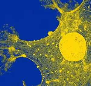
Cytoskeleton, TEM
Cytoskeleton. Coloured transmission electron micrograph (TEM) of the cytoskeleton of a human skin cell. The cell nucleus is at centre right

Background imageCell Biology Collection: Protein, microtubules and cell, artwork

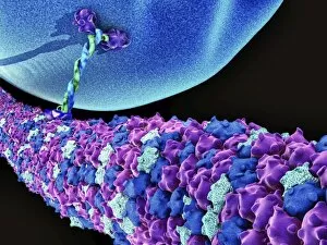
Protein, microtubules and cell, artwork
3D computer artwork of a protein attached to microtubules, transporting vesicles and other important parts of the cell. Microtubules are polymers of the protein tubulin

Background imageCell Biology Collection: Stem cell therapy, artwork

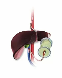
Stem cell therapy, artwork
Stem cell therapy. Artwork showing stem cells (in green cylinder) being added to a patients bloodstream in the region of the liver (brown)

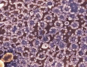
Background imageCell Biology Collection: Cytoskeleton, SEM

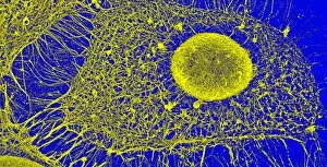
Cytoskeleton, SEM
Cytoskeleton. Coloured scanning electron micrograph (SEM) of the cytoskeleton of a human skin cell. The cell nucleus is oval. The rest of the cells contents have been biochemically extracted

Background imageCell Biology Collection: Keratinocyte skin cells, light micrograph

Keratinocyte skin cells, light micrograph
Keratinocyte skin cells. Fluorescent light micrograph of the cytoskeleton of human keratinocyte skin cells. Cell nuclei are oval. The rest of the cells contents have been biochemically extracted

Background imageCell Biology Collection: Retinal rod cell anatomy, diagram

Retinal rod cell anatomy, diagram

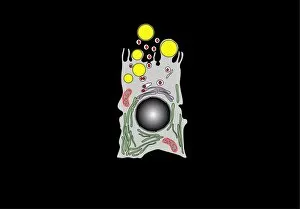
Background imageCell Biology Collection: Milk-producing cell, artwork

Milk-producing cell, artwork
Milk-producing cell. Artwork of the anatomical structure of a lacteal (milk-producing) cell. Normal cell organelles include the nucleus (black, centre), the golgi apparatus (white)

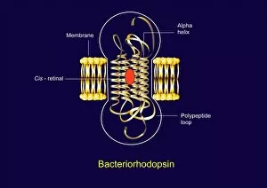
Background imageCell Biology Collection: Bacteriorhodopsin, diagram

Bacteriorhodopsin, diagram
Bacteriorhodopsin. Diagram of the molecular structure of bacteriorhodopsin, a protein found in primitive micro-organisms known as Archaea

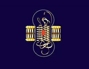
Background imageCell Biology Collection: Bacteriorhodopsin, artwork

Bacteriorhodopsin, artwork
Bacteriorhodopsin. Artwork of the molecular structure of bacteriorhodopsin, a protein found in primitive micro-organisms known as Archaea
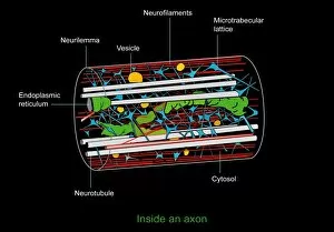
Background imageCell Biology Collection: Axon anatomy, diagram

Axon anatomy, diagram
Axon anatomy. Diagram of the anatomical structure of an axon, the main extension (dendrite) of a nerve cell. Cellular components include endoplasmic reticulum (green), neurotubules (white)

Background imageCell Biology Collection: Capillary, artwork

Capillary, artwork
Capillary. Artwork showing the anatomical structure of a capillary seen in cross-section

Background imageCell Biology Collection: Retinal rod cell anatomy, artwork

Retinal rod cell anatomy, artwork
Retinal rod cell anatomy. Artwork of the external (left) and internal (right) anatomical structure of a retinal cell. These cells are found in the retina that lines the back of the eyes

Background imageCell Biology Collection: Milk-producing cell, diagram

Milk-producing cell, diagram
Milk-producing cell. Diagram of the anatomical structure of a lacteal (milk-producing) cell. Normal cell organelles include the nucleus (black, centre), the golgi apparatus (white)

Background imageCell Biology Collection: Animal cell anatomy, artwork

Animal cell anatomy, artwork
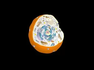
Animal cell anatomy. Artwork showing the internal and external anatomy of an animal cell

Background imageCell Biology Collection: Macrophage engulfing pathogen, artwork

Macrophage engulfing pathogen, artwork
Macrophage engulfing pathogen. Artwork of a macrophage cell (pink) engulfing a pathogen (green), a process known as phagocytosis. The pathogen will be destroyed once it has been engulfed

Background imageCell Biology Collection: Activated platelets, artwork

Activated platelets, artwork
Activated platelets. Artwork of platelets (thrombocytes) that have been activated. Platelets are part of the blood. When a blood vessel is damaged

Background imageCell Biology Collection: Macrophage engulfing pathogens, artwork

Macrophage engulfing pathogens, artwork
Macrophage engulfing pathogens. Artwork of a macrophage cell (pink) engulfing pathogens (green), a process known as phagocytosis. The pathogens will be destroyed once they have been engulfed

Background imageCell Biology Collection: Damaged cardiac tissue, artwork

Damaged cardiac tissue, artwork
Damaged cardiac tissue. Artwork of damaged heart fibres and cells in heart muscle tissue (myocardium). This can occur due to reduced blood supply (ischaemia)

Background imageCell Biology Collection: mRNA leaving the nucleus, artwork

mRNA leaving the nucleus, artwork. mRNA (messenger ribonucleic acid, orange) is the intermediary molecule between DNA (deoxyribonucleic acid) and its protein product

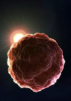
Background imageCell Biology Collection: Stem cell, artwork

Stem cell, artwork
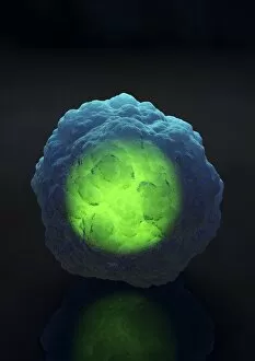
Stem cell with a glowing nucleus, computer artwork. A stem cell is an undifferentiated cell that can produce other types of cell when it divides

Background imageCell Biology Collection: Animal cell, artwork

Animal cell, artwork
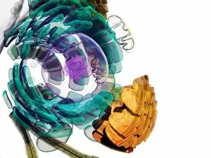
Animal cell. Computer artwork of an expanded animal cell. At centre left is the nucleus (purple sphere), which contains the cells genetic information in the form of DNA (deoxyribonucleic acid)

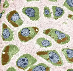
Background imageCell Biology Collection: Cartilage cells, TEM

Cartilage cells, TEM
Cartilage cells. Coloured transmission electron micrograph (TEM) of a section through chondrocytes from nasal hyaline cartilage

Background imageCell Biology Collection: Stem cell research, conceptual artwork

Stem cell research, conceptual artwork
Stem cell research, conceptual computer artwork. The glowing light behind the stem cell represents the dawning of a new era of medicine involving stem cells

Background imageCell Biology Collection: Spleen cell, TEM

Spleen cell, TEM
Spleen cell. Coloured transmission electron micrograph (TEM) of a section through a cell in the spleen, an organ located in the upper left side of the abdomen in vertebrate animals

Background imageCell Biology Collection: Spleen cells, TEM

Spleen cells, TEM
Spleen cells. Coloured transmission electron micrograph (TEM) of a section through cells in the spleen, an organ located in the upper left side of the abdomen in vertebrate animals

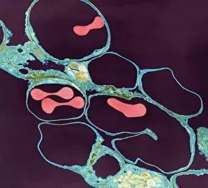
Background imageCell Biology Collection: Lung alveoli and red blood cells, TEM

Lung alveoli and red blood cells, TEM
Lung alveoli and red blood cells. Coloured transmission electron micrograph (TEM) of a section through lung alveoli (blue) and red blood cells (red)

Background imageCell Biology Collection: Mitochondrial energy, conceptual image

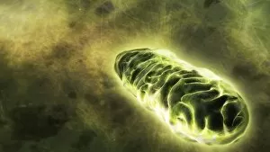
Mitochondrial energy, conceptual image. Computer artwork of a glowing mitochondrion, representing the energy produced by these cell organelles

Background imageCell Biology Collection: Heart muscle cell, SEM

Heart muscle cell, SEM
Heart muscle cell. Coloured scanning electron micrograph (SEM) of a heart muscle cell (cardiac myocyte). Cardiac muscle is a specialised type of striated muscle found in the heart

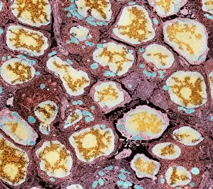
Background imageCell Biology Collection: Spinal root nerves, SEM

Spinal root nerves, SEM
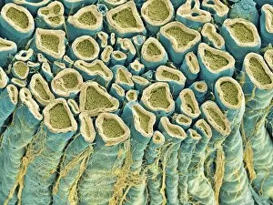
Spinal root nerves. Coloured scanning electron micrograph (SEM) of a sectioned spinal root nerve bundle, showing individual axons, extensions of nerve cells)

Background imageCell Biology Collection: Lung alveoli and blood cells, TEM

Lung alveoli and blood cells, TEM
Lung alveoli and blood cells. Coloured transmission electron micrograph (TEM) of a section through lung alveoli and blood cells

Background imageCell Biology Collection: Hippocampus neuron, TEM

Hippocampus neuron, TEM
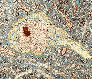
Hippocampus neuron. Coloured transmission electron micrograph (TEM) of a section through a neuron from the hippocampus, a structure in the brain responsible for long-term memory

Background imageCell Biology Collection: Nucleolus, SEM

Nucleolus, SEM
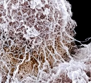
Nucleolus, coloured scanning electron micrograph (SEM). The nucleolus is responsible for producing components of ribosomes, the cells protein-manufacturing organelles

Background imageCell Biology Collection: Nuclear pore complexes, SEM

Nuclear pore complexes, SEM
Nuclear pore complexes. Coloured scanning electron micrograph (SEM) of the surface of a cell nucleus showing the numerous nuclear pore complexes (NPCs, rings) in its envelope

Background imageCell Biology Collection: Blood cells in blood vessel, artwork

Blood cells in blood vessel, artwork
Blood cells in blood vessel, computer artwork. Red blood cells (erythrocytes, red) distribute oxygen to body tissues, and carry waste carbon dioxide back to the lungs

Background imageCell Biology Collection: Red blood cells in blood vessel, artwork

Red blood cells in blood vessel, artwork
Red blood cells in blood vessel, computer artwork. Red blood cells (erythrocytes) are biconcave, giving them a large surface area for gas exchange, and highly elastic

Background imageCell Biology Collection: Plant cell, SEM

Plant cell, SEM
Plant cell. Coloured scanning electron micrograph (SEM) of a section through a plant cell, revealing its internal structure. The cell is encased in a cellulose, hemicellulose and pectin cell wall

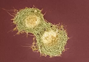
Background imageCell Biology Collection: Cell division, SEM

Cell division, SEM
Cell division. Coloured scanning electron micrograph (SEM) of two animal cells during cytokinesis (cell division). Cytokinesis occurs after nuclear division (mitosis)

Background imageCell Biology Collection: Red blood cells, artwork

Red blood cells, artwork
Red blood cells, computer artwork. Red blood cells (erythrocytes) are biconcave, giving them a large surface area for gas exchange, and highly elastic


For sale as Licensed Images

Choose your image, Select your licence and Download the media

"Exploring the Intricacies of Cell Biology: A Fascinating Journey into Cellular Life" Nerve and glial cells, captured through a mesmerizing light micrograph, reveal the intricate network that supports our nervous system. T lymphocytes battling cancer cells - witness this epic battle in stunning detail with SEM C001 / 1679 imagery. Anaesthetic inhibiting an ion channel - delve into the world of cellular communication as we uncover how anaesthetics affect these vital channels (C015 / 6718). Glial cells take center stage in a captivating confocal light micrograph, showcasing their crucial role in supporting neurons. Witness the beauty of glial stem cell culture under a microscope; it's like exploring an enchanting galaxy within our own bodies (light micrograph). Dendritic cells come to life through exquisite artwork, highlighting their pivotal role in immune response and defense mechanisms. Pine pollen grains unveil their intricate structure under a light microscope – nature's tiny marvels never cease to amaze us. Dive deep into the inner workings of pine stems with a captivating light micrograph – discover the building blocks that support these majestic trees. Explore the hidden wonders of lime tree stems through a mesmerizing light micrograph – witness nature's architectural masterpiece up close. Uncover various cell types brought to life through breathtaking artwork – each one playing its unique part in maintaining life's delicate balance. Immerse yourself in neural stem cell culture and witness firsthand how these remarkable cells hold immense potential for regenerative medicine. The artistic portrayal of cell membranes (C013 / 7467) invites you to ponder upon their crucial role as gatekeepers controlling molecular traffic within living organisms. Embark on this awe-inspiring journey into cell biology and unlock the secrets that shape all living beings from within.