
Cell Culture Collection
"Unlocking the Secrets of Cell Culture: Exploring the Fascinating World Within" Immerse yourself in the captivating realm of cell culture


For sale as Licensed Images
Choose your image, Select your licence and Download the media
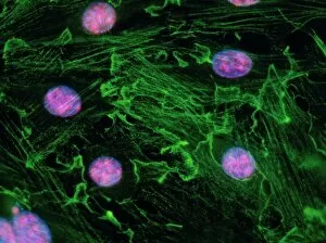
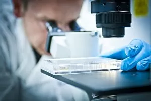
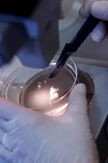
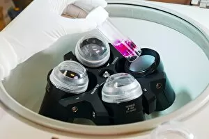
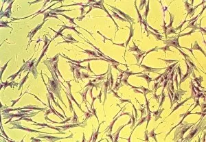
"Unlocking the Secrets of Cell Culture: Exploring the Fascinating World Within" Immerse yourself in the captivating realm of cell culture, where scientists delve into microscopic wonders that hold immense potential for scientific breakthroughs. In this image-rich journey, we embark on a visual exploration of various facets within this intricate field. Our first stop takes us to an immunofluorescent light micrograph showcasing fibroblast cell nuclei. These vibrant clusters represent the building blocks of life, offering insights into cellular behavior and functions. Moving forward, we encounter a petri plate brimming with cells undergoing cultivation. This essential tool provides researchers with a controlled environment to nurture and study these delicate organisms as they grow and multiply. Next up is an inverted light micrograph revealing MDCK cells – another vital component in cell culture research. Through meticulous observation under a microscope, scientists unravel their characteristics and behaviors, paving the way for advancements in medical science. Speaking of microscopes, our journey wouldn't be complete without glimpses of dedicated researchers peering through these powerful lenses. Their unwavering dedication allows them to uncover hidden truths within each specimen they examine. As we venture deeper into this world, we come across carbon dioxide incubators – indispensable equipment ensuring optimal growth conditions for cultured cells. These controlled environments mimic natural habitats while providing necessary nutrients and gaseous requirements for thriving cultures. The images captured from different angles highlight how these incubators play an integral role in maintaining stable conditions crucial for successful experiments and discoveries. The synchronized dance between technology and biology unfolds before our eyes. Amongst all these marvels lies yet another wonder - cultured nerve cells pulsating with vitality. These specialized cells offer invaluable insights into neurological disorders such as Alzheimer's or Parkinson's disease—fueling hope for future treatments or even cures. Finally returning full circle to our starting point—an immunofluorescent LM reveals fibroblast cell nuclei once again—a reminder that cell culture is an ever-evolving field.