Cell Division Collection (#4)

"Unlocking the Mysteries of Life: Exploring Cell Division Through Micrographs and Illustrations" Witness the intricate dance of life as cells divide through mitosis

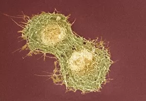
Background imageCell Division Collection: Cell division, SEM

Cell division, SEM
Cell division. Coloured scanning electron micrograph (SEM) of two animal cells during cytokinesis (cell division). Cytokinesis occurs after nuclear division (mitosis)

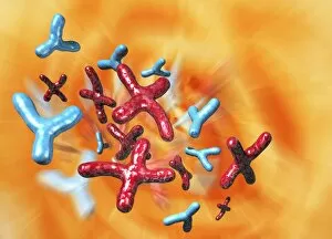
Background imageCell Division Collection: X and Y chromosomes, artwork

X and Y chromosomes, artwork
X and Y chromosomes. Computer artwork of X (red) and Y (blue) chromosomes. Chromosomes are a packaged form of the genetic material DNA (deoxyribonucleic acid)

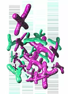
Background imageCell Division Collection: Chromosomes and DNA, artwork

Chromosomes and DNA, artwork
Chromosomes and DNA. Computer artwork of X (pink) and Y (green) chromosomes with an autoradiogram in the background. Chromosomes are a packaged form of DNA (deoxyribonucleic acid)

Background imageCell Division Collection: Young stem, light micrograph

Young stem, light micrograph. Longitudinal section through a young stem showing an intercalary meristem. Intercalary meristems are found at the base of grass leaves

Background imageCell Division Collection: Chromosome, artwork

Chromosome, artwork
Chromosome, computer artwork. Chromosomes are a packaged form of the genetic material DNA (deoxyribonucleic acid, blue strands)

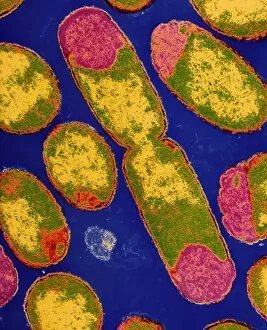
Background imageCell Division Collection: TEM of dividing E. coli bacterium

TEM of dividing E. coli bacterium
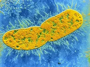
False-colour transmission electron micrograph of the bacterium Escherichia coli, a normal inhabitant of the human intestine

Background imageCell Division Collection: Klebsiella pneumoniae bacterium

Klebsiella pneumoniae bacterium
False colour transmission electron micrograph (shadow technique) of Klebsiella pneumoniae, a rodlike bacterium associated with pneumonia & other respiratory infections in humans

Background imageCell Division Collection: Cell nuclei with chromatin

Cell nuclei with chromatin
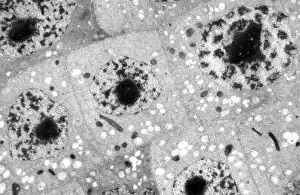
Transmission electron micrograph of several nuclei in meristematic tissue of the maize plant Zea mays root tip at interphase. Large nucleus approaching mitosis showing clumping of chromatin

Background imageCell Division Collection: Stenotrophomonas sp. bacteria, TEM

Stenotrophomonas sp. bacteria, TEM
Stenotrophomonas maltophilia bacteria. Coloured transmission electron micrograph (TEM) of a sect- ion through Stenotrophomonas maltophilia bacteria. They are seen on a fractal background

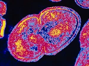
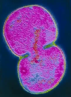
Background imageCell Division Collection: Meningitis bacteria dividing

Meningitis bacteria dividing
Neisseria meningitidis. Coloured Transmission electron micrograph of the bacteria Neisseria meningitidis, the agent which causes meningitis

Background imageCell Division Collection: Mycobacterium dividing, TEM

Mycobacterium dividing, TEM
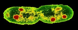
Mycobacterium dividing. Coloured transmission electron micrograph (TEM) of a Mycobacterium sp. bacterium undergoing binary fission

Background imageCell Division Collection: Coloured TEM of Salmonella bacteria

Coloured TEM of Salmonella bacteria
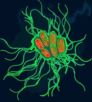
Salmonella sp. Coloured transmission electron micrograph (TEM) of a cluster of Salmonella sp. bacteria. The cells are rod-shaped with long hair- like flagellae

Background imageCell Division Collection: Coloured TEM of Enterococcus faecalis

Coloured TEM of Enterococcus faecalis
Enterococcus faecalis. Coloured transmission electron micrograph (TEM) of Enterococcus faecalis bacteria (previously known as Streptococcus faecalis)

Background imageCell Division Collection: Dividing Streptococcus sp. bacterium

Dividing Streptococcus sp. bacterium
False-colour transmission electron micrograph (TEM) of the Gram-positive bacterium Streptococcus sp.; the elongated cell at top is in the process of division (replication)

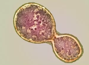
Background imageCell Division Collection: Cryptococcus cell dividing, TEM

Cryptococcus cell dividing, TEM
Cryptococcus cell dividing. Coloured transmission electron micrograph (TEM) of the asexual budding (lower right) of a Cyptococcus sp. yeast cell

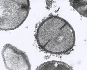
Background imageCell Division Collection: TEM of Staphylococcus aureus bacteria

TEM of Staphylococcus aureus bacteria
Staphylococcus bacteria. Transmission electron micrograph (TEM) of sectioned Staphylococcus aureus bacteria. Staphylococcus aureus are Gram- positive coccal (spherical) bacteria

Background imageCell Division Collection: Arginine molecule

Arginine molecule. Computer model of a molecule of the amino acid arginine. Atoms are represented as rods and are colour-coded: carbon (green), hydrogen (white), oxygen (red) and nitrogen (blue)

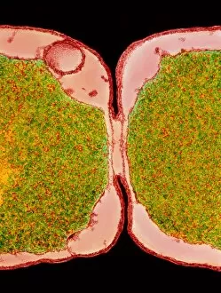
Background imageCell Division Collection: Coloured TEM of E. coli bacteria dividing

Coloured TEM of E. coli bacteria dividing
E. Coli bacteria dividing. Transmission electron micrograph of the division zone between two new Escherichia coli bacteria showing the cells undergoing binary fission

Background imageCell Division Collection: Moraxella catarrhalis bacterium dividing

Moraxella catarrhalis bacterium dividing

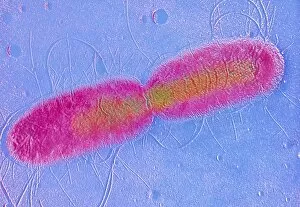
Background imageCell Division Collection: F / col shadow TEM of E. coli dividing

F / col shadow TEM of E. coli dividing
False-colour transmission electron micrograph (TEM) of the bacterium Escherichia coli, a normal inhabitant of the human intestine, in the act of dividing

Background imageCell Division Collection: Cell division in Salmonella bacterium

Cell division in Salmonella bacterium
Salmonella cell dividing. Coloured Transmission electron micrograph of the bacterium Salmonella typhimurium undergoing division by binary fission

Background imageCell Division Collection: TEM E. Coli

TEM E. Coli
False colour transmission electron micrograph of the bacterium Escherichia coli, a normal member of human intestinal flora

Background imageCell Division Collection: Dividing cell in maize root tip

Dividing cell in maize root tip

Background imageCell Division Collection: TEM of E. coli bacterium

TEM of E. coli bacterium
False colour transmission electron micrograph (shadow technique) of the bacterium Escherichia coli, a normal inhabitant of the human intestine

Background imageCell Division Collection: Staphylococcus aureus bacteria dividing

Staphylococcus aureus bacteria dividing
Staphylococcus bacteria. Coloured transmission electron micrograph (TEM) of Staphylococcus aureus bacteria (light blue), some undergoing cell division

Background imageCell Division Collection: Cyclin-dependent kinase 2 enzyme

Cyclin-dependent kinase 2 enzyme, molecular model. This enzyme is found in cells, where it is involved in regulating the cell cycle, the cycle of cell division and cell growth


For sale as Licensed Images

Choose your image, Select your licence and Download the media

"Unlocking the Mysteries of Life: Exploring Cell Division Through Micrographs and Illustrations" Witness the intricate dance of life as cells divide through mitosis, captured in a mesmerizing light micrograph. Delve into the microscopic world of E. Coli bacteria, where a constant process, revealed in a fluorescent micrograph. Marvel at the astonishing progress of embryo development just 24-36 hours after fertilization, depicted in another captivating fluorescent micrograph. Explore the inner workings of an E. Coli bacterium under high magnification with a TEM image that showcases its dividing nature. Immerse yourself in the vibrant false-color TEM image that unveils the beauty within an E. coli bacterium during division. Observe with awe as an E. coli bacterium undergoes division, revealing its remarkable ability to multiply and thrive. Journey into human biology as SEM captures striking images of chromosomes during cell division – a crucial process for growth and repair. Uncover the intricacies of cellular reproduction through SEM imagery that highlights various stages across different organisms. Step back in time with a fascinating 19th-century medical illustration showcasing facial nerves involved in complex processes like cell division. Explore nuclear division through an illuminating illustration that unravels how genetic material is precisely distributed during this vital cellular event. In this captivating collection, we embark on an enlightening journey through various scientific techniques and illustrations to unravel one of life's most fundamental processes – cell division.