Cells Collection (#77)

"Exploring the Intricacies of Cells: From Cerebellum Tissue to Geranium Anther" Delving into the complexity of cells

Background imageCells Collection: Light Micrograph (LM): Cells from a hair on the stamen of the common spiderwort (Tradescantia)

Light Micrograph (LM): Cells from a hair on the stamen of the common spiderwort (Tradescantia)
LRDS-171 Light Micrograph (LM): Cells from a hair on the stamen of the common spiderwort (Tradescantia) Tradescantia sp David Spears (Last Refuge)

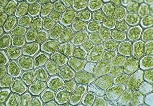
Background imageCells Collection: Light Micrograph (LM): The cellular struture of a liverwort plant (Hepatica)

Light Micrograph (LM): The cellular struture of a liverwort plant (Hepatica); Magnification x1200 (on 10)
LRDS-190 Light Micrograph (LM): The cellular struture of a liverwort plant (Hepatica) Magnification x1200 (on 10.5 cm width print) Hepatica sp David Spears (Last Refuge)

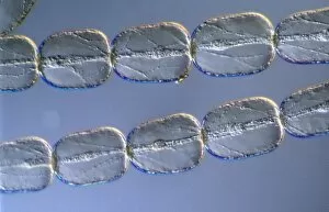
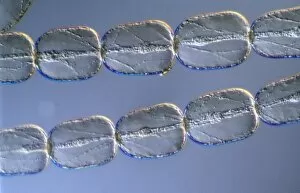
Background imageCells Collection: Light Micrograph (LM): A single cell from a hair on the stamen of the common spiderwort

Light Micrograph (LM): A single cell from a hair on the stamen of the common spiderwort (Tradescantia)
LRDS-170 Light Micrograph (LM): A single cell from a hair on the stamen of the common spiderwort (Tradescantia) Tradescantia sp David Spears (Last Refuge)

Background imageCells Collection: Light Micrograph (LM) of a plant cell chloroplasts, the site where photosynthesis takes place

Light Micrograph (LM) of a plant cell chloroplasts, the site where photosynthesis takes place

Background imageCells Collection: Light Micrograph (LM) of cells from a hair on the stamen of the common spiderwort

Light Micrograph (LM) of cells from a hair on the stamen of the common spiderwort (Tradescantia)

Background imageCells Collection: Light Micrograph (LM) of onion skin cells, magnification x 600

Light Micrograph (LM) of onion skin cells, magnification x 600

Background imageCells Collection: Light Micrograph (LM) of the cellular structure of the non-vascular plant liverwort

Light Micrograph (LM) of the cellular structure of the non-vascular plant liverwort (Hepatica) showing chloroplasts and oil bodies, magnification x1200

Background imageCells Collection: Light Micrograph (LM) of a concentration of cells on the epidermis of a plant showing stomata

Light Micrograph (LM) of a concentration of cells on the epidermis of a plant showing stomata

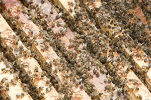
Background imageCells Collection: A bee that has deformed wings as a result of attack by Varoa mites in a beehive in Cockermouth

A bee that has deformed wings as a result of attack by Varoa mites in a beehive in Cockermouth, Cumbria
Ashley Cooper / SpecialistStock

Background imageCells Collection: Apistan being used to combat the Vaoa mite in hives in Cockermouth Cumbria, UK

Apistan being used to combat the Vaoa mite in hives in Cockermouth Cumbria, UK
Ashley Cooper / SpecialistStock

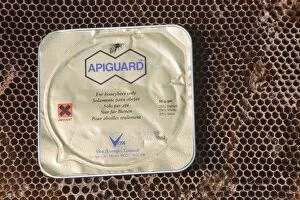
Background imageCells Collection: Apiguard being used to combat the Vaoa mite in hives in Cockermouth Cumbria, UK

Apiguard being used to combat the Vaoa mite in hives in Cockermouth Cumbria, UK
Ashley Cooper / SpecialistStock

Background imageCells Collection: A beehive in Cockermouth, Cumbria, UK that has been infected and damaged by the Varoa mite

A beehive in Cockermouth, Cumbria, UK that has been infected and damaged by the Varoa mite
Ashley Cooper / SpecialistStock

Background imageCells Collection: Bill Mackereth, a beekeeper from Cockermouth, Cumbria, UK, checks his hives for signs of Varoa

Bill Mackereth, a beekeeper from Cockermouth, Cumbria, UK, checks his hives for signs of Varoa mite damage
Ashley Cooper / SpecialistStock

Background imageCells Collection: Frogspawn

Frogspawn
Ashley Cooper / SpecialistStock

Background imageCells Collection: Tropical rainforest on Fiji

Tropical rainforest on Fiji
Ashley Cooper / SpecialistStock

Background imageCells Collection: Tropical rainforest plants on Fiji

Tropical rainforest plants on Fiji
Ashley Cooper / SpecialistStock

Background imageCells Collection: Light Micrograph (LM): Onion skin cells; Magnification x600 (on 10. 5 cm width print)

Light Micrograph (LM): Onion skin cells; Magnification x600 (on 10. 5 cm width print)
LRDS-252 Light Micrograph (LM): Onion skin cells; Magnification x600 (on 10.5 cm width print) David Spears (Last Refuge) / ardea.com Last Refuge Please note that prints are for personal display

Background imageCells Collection: Light Micrograph (LM): A concentration of cells on the epidermis of a plant showing stomata

Light Micrograph (LM): A concentration of cells on the epidermis of a plant showing stomata
LRDS-197 Light Micrograph (LM): A concentration of cells on the epidermis of a plant showing stomata David Spears (Last Refuge)

Background imageCells Collection: Russia / 1936 / Prisoners

Russia / 1936 / Prisoners
Political prisoners are confined in brightly lit cells in temperatures of 40 degrees centigrade, and given salt water to drink, so they are driven mad

Background imageCells Collection: Refuelling in Space

Refuelling in Space
Recharging the Energy Cells of a Future Aircraft in Mid- Flight

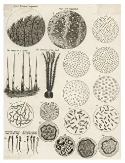
Background imageCells Collection: Microscopic Objects

Microscopic Objects
A variety of living and non- living objects magnified through a microscope

Background imageCells Collection: Battery constructed at the Royal Institution, London

Battery constructed at the Royal Institution, London
A large battery constructed at the Royal Institution, London, by Wollaston, for Davy

Background imageCells Collection: Construction of a voltaic battery in Paris

Construction of a voltaic battery in Paris
Construction of a large voltaic battery at the Ecole Polytechnique in Paris, covering 54 square metres, on the orders of Napoleon


For sale as Licensed Images

Choose your image, Select your licence and Download the media

"Exploring the Intricacies of Cells: From Cerebellum Tissue to Geranium Anther" Delving into the complexity of cells, we examine cerebellum tissue through a captivating light micrograph. Behind the walls of Wandsworth Prison in southwest London, cells silently carry out their vital functions within inmates' bodies. Witnessing the intricate network of nerve and glial cells, a mesmerizing light micrograph reveals their interconnectedness. At the synapse nerve junction, captured by TEM imaging, we witness the fascinating communication between cells that allows our body to function seamlessly. Within Pentonville Prison in Islington, North London, hidden stories unfold as various cell types coexist amidst confinement. Shedding light on groundbreaking research, T lymphocytes and cancer they can magnified under SEM imaging - offering hope for future treatments (SEM C001 / 1679). Peering into hippocampus brain tissue unveils a world where countless cells work together to shape our memories and emotions. Glial cells take center stage in a confocal light micrograph - showcasing their crucial role in supporting and protecting neurons within our nervous system. HeLa cells come alive under scrutiny with an intriguing light micrograph (C017 / 8299), reminding us of Henrietta Lacks' immortal contribution to medical science. Journey back in time as Aylesbury Prison's historical significance intertwines with its cellular inhabitants from 1900 onwards. Exploring beyond human boundaries, dicotyledon plant stems reveal their cellular architecture through an enchanting light micrograph. Zooming closer than ever before, geranium anthers expose their delicate beauty under SEM imaging - unveiling nature's remarkable cellular structures.