
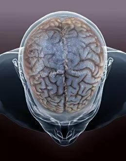
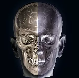
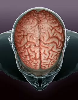
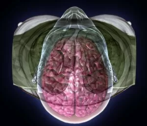
Cerebral Hemisphere Collection
The cerebral hemisphere, a fascinating part of the normal human brain, can be explored through various imaging techniques such as MRI and CT scans


For sale as Licensed Images
Choose your image, Select your licence and Download the media
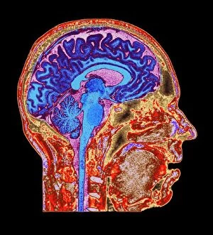
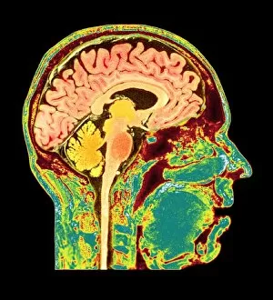
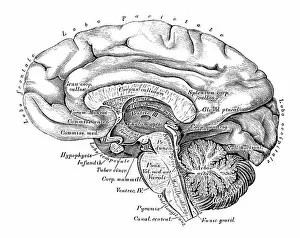
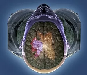
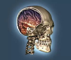
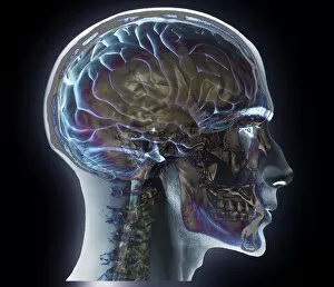
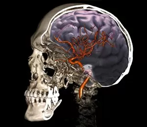
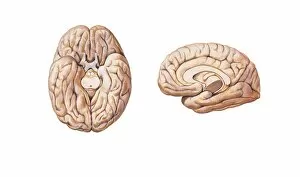
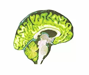
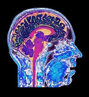
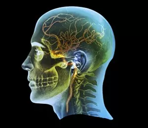
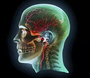
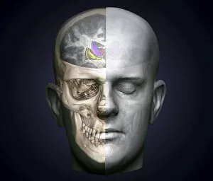
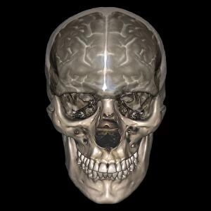
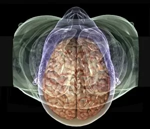
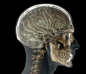
The cerebral hemisphere, a fascinating part of the normal human brain, can be explored through various imaging techniques such as MRI and CT scans. In MRI scan C016 / 8845 and C016 / 8850, we get an inside look at the intricate details of this vital organ. Meanwhile, artwork depicting brain anatomy provides us with a visual representation of its complexity. Scientific illustrations showcasing the side view of the human brain (C016 / 8843) allow us to delve deeper into understanding its structure. Additionally, in the 3D CT scan C016 / 6333, we witness a comprehensive view of both the normal skull and brain. However, not all images portray healthy conditions; stroke is captured in MRI and 3D CT scans (C016 /6419), reminding us of the importance of maintaining optimal brain health. Expanding our exploration beyond just cerebral hemispheres, other scans like those for normal head and neck (C016/6337) or external carotid artery (C016/6341) provide valuable insights into related areas that contribute to overall neurological well-being. Whether it's through medical imaging or scientific illustrations, studying brain anatomy grants us invaluable knowledge about cerebral hemispheres. From their upper lateral surface to their lower and medial surfaces - every detail counts when unraveling the mysteries within our minds.