
Cervical Vertebrae Collection
The cervical vertebrae, also known as the neck vertebrae, play a crucial role in supporting and protecting the upper body


For sale as Licensed Images
Choose your image, Select your licence and Download the media
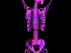
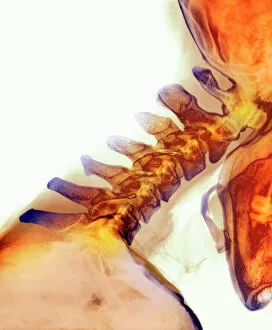
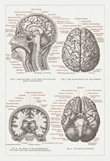
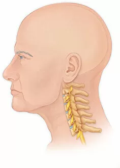
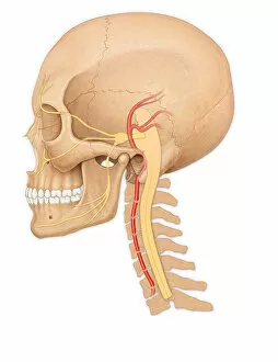
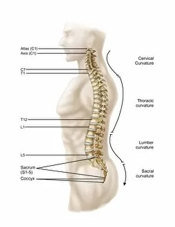
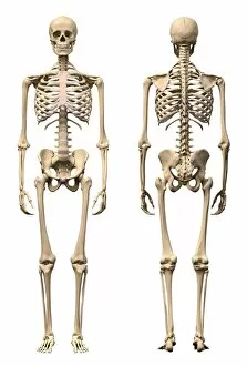
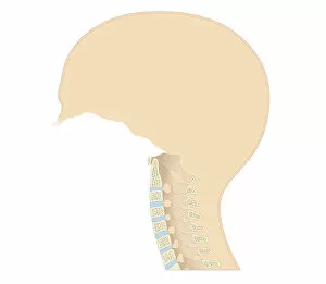
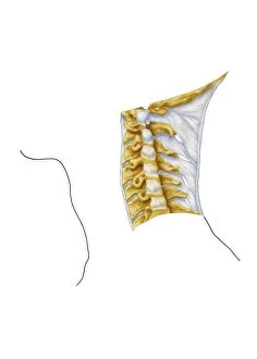
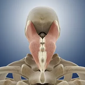
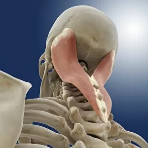
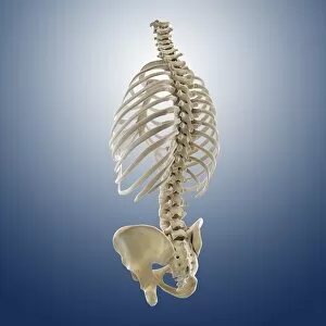
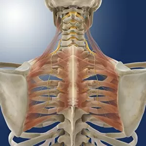
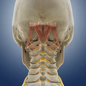
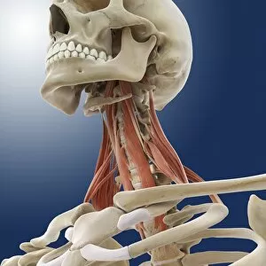
The cervical vertebrae, also known as the neck vertebrae, play a crucial role in supporting and protecting the upper body. In an X-ray image, these bones can be seen extending from the base of the skull down to the top of the thoracic spine. When depicted in computer artwork showcasing the entire upper body skeleton, their significance becomes evident. Dating back to 1876, a lithograph provides an intricate view of human brain anatomy alongside these cervical vertebrae. Meanwhile, another illustration from 1866 focuses solely on dissecting and understanding their structure. From different angles and perspectives, various images shed light on this vital part of our skeletal system. A normal lateral view showcases both a man's head and neck with his skull intact while highlighting how they connect to form a healthy spinal cord. Similarly, a side view emphasizes how these vertebrae align within an adult skull. To truly grasp their complexity and function within our bodies, it is essential to explore related elements such as neck muscles and nerves or suboccipital muscles and nerves – all beautifully depicted through detailed artwork. When examining medical imagery like X-rays or cross-sectional biomedical illustrations that zoom in on specific areas like the cervical curve or individual cervical vertebrae themselves – we gain insight into potential issues that may arise if not properly cared for. Ultimately, whether viewed through historical lithographs or modern-day digital renderings - studying the anatomy allows us to appreciate their integral role in maintaining a healthy spine and overall well-being.