
Chromatid Collection (#2)
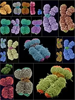
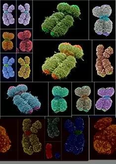
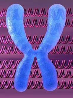
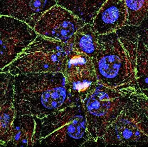
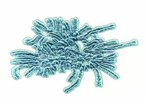
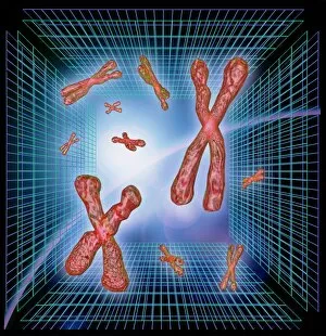
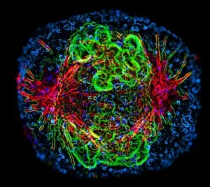
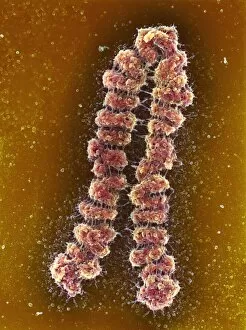
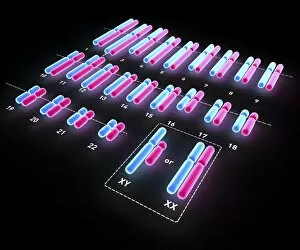
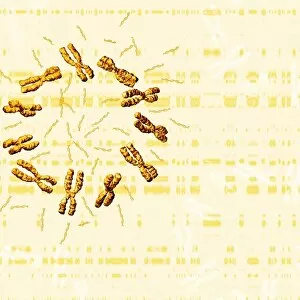
Chromatids are the fascinating structures that make up chromosomes, including the iconic X and Y chromosomes


For sale as Licensed Images
Choose your image, Select your licence and Download the media
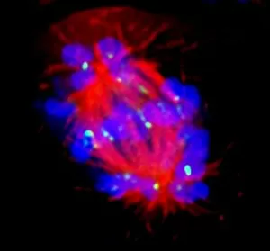
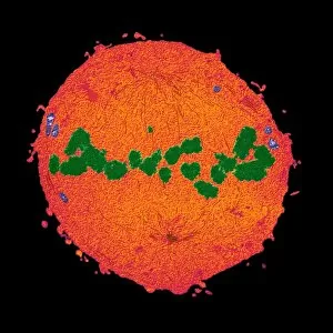
Chromatids are the fascinating structures that make up chromosomes, including the iconic X and Y chromosomes. In the intricate process of mitosis, these chromatids play a crucial role in cell division. When observed under a light micrograph, dividing cells reveal their mesmerizing beauty as chromatids align themselves perfectly to ensure accurate distribution of genetic material. The X and Y chromosomes hold special significance as they determine an individual's biological sex. These distinct chromosome pairs can be visualized through scanning electron microscopy (SEM), showcasing their unique features and patterns. An illustration of a human chromosome provides further insight into its structure, highlighting essential components such as the chromatid, centromere, short arm, and long arm. SEM images continue to unveil the astonishing complexity of human chromosomes with their intricate arrangements. Studying chromatids not only deepens our understanding of genetics but also sheds light on various aspects of life itself. From inheritance patterns to genetic disorders, these tiny entities hold vast amounts of information that shape who we are as individuals. As we delve deeper into the microscopic world of cells and chromosomes, SEM images offer us glimpses into this hidden realm. The striking visuals captured by this technique allow scientists to explore chromosomal abnormalities or anomalies that may arise during development or contribute to certain diseases. In essence they can more than just thread-like structures within our cells; they represent the blueprint for life itself. Through ongoing research and technological advancements like SEM imaging techniques, we continue unraveling their mysteries while marveling at nature's incredible design.