
Circulatory System Collection (#7)
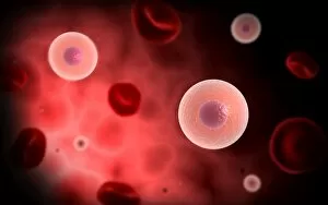
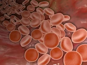
The circulatory system is a complex network of blood vessels that ensures the proper functioning of our bodies


For sale as Licensed Images
Choose your image, Select your licence and Download the media
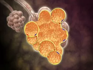
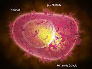
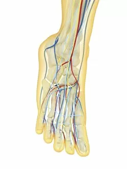
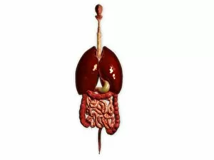
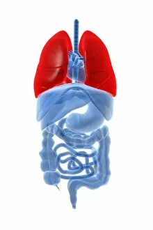
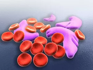
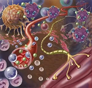
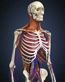
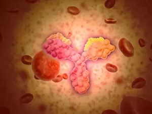
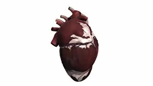
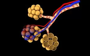
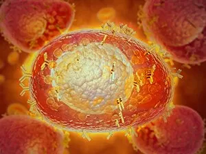
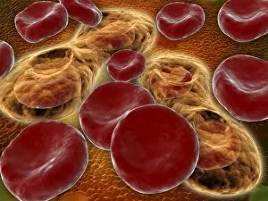
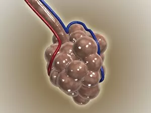
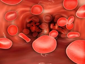
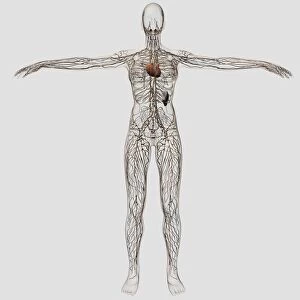
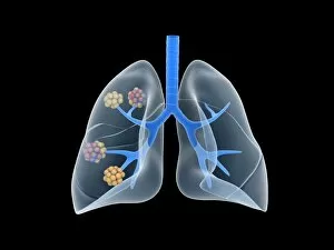
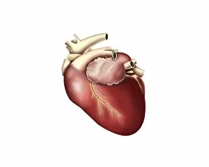
The circulatory system is a complex network of blood vessels that ensures the proper functioning of our bodies. From the brain blood vessels to the arterial system, this intricate system keeps us alive and thriving. In 3D angiogram C007 / 1981, we get a glimpse into the fascinating world of brain blood vessels. These delicate pathways supply oxygen and nutrients to our most vital organ, allowing it to function at its best. The heart, often referred to as the "engine" of our bodies, plays a crucial role in pumping oxygenated blood throughout our systems. Human heart anatomy artwork showcases its intricate structure and highlights its importance in sustaining life. Dating back to the 18th century, an illustration of the arterial system reminds us of how far medical knowledge has come. This detailed depiction shows how arteries branch out from larger vessels like tree roots, ensuring every part of our body receives essential nutrients. X-ray images reveal another aspect of circulation - neck and shoulder arteries. These powerful images allow us to see these vital pathways up close and appreciate their role in supplying oxygen-rich blood to these areas. Artwork from 1825 takes us on a journey through male groin arteries. While it may seem unusual today, this historical piece demonstrates early attempts at understanding human anatomy and circulation. A diagram showing bones, veins, and arteries in a human arm and hand provides insight into how circulation reaches even our extremities. It's incredible to think about how each part works together seamlessly for optimal functionality. Blood coagulation cascade artwork captures one critical aspect: clotting mechanisms that prevent excessive bleeding when injuries occur. This intricate process involves various proteins working harmoniously within our circulatory system. Anatomical artwork depicting arm circulation offers yet another perspective on this vast network within us all. Understanding how blood flows through different regions helps comprehend overall health better while appreciating the complexity involved. Heart and lungs work hand-in-hand as they ensure proper oxygenation of our blood.