
Coagulated Collection
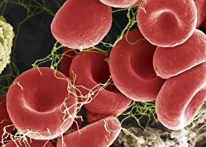
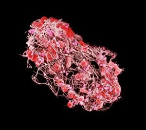
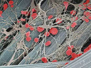
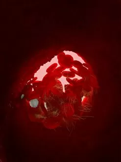
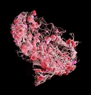
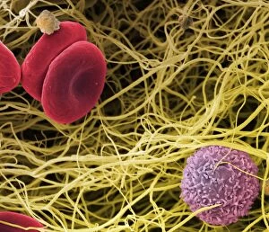
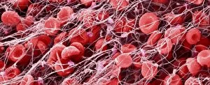
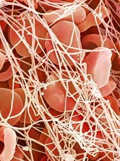
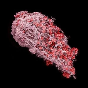
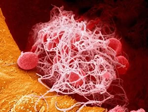
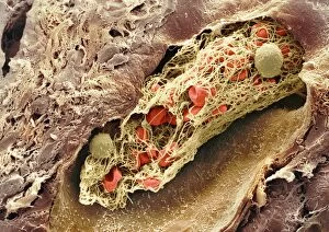
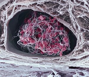
"Coagulated: Unveiling the Intricate World of Blood Clots" Step into the microscopic realm and witness the fascinating intricacies of coagulation


For sale as Licensed Images
Choose your image, Select your licence and Download the media
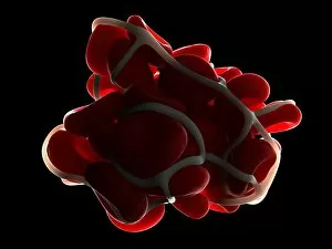
"Coagulated: Unveiling the Intricate World of Blood Clots" Step into the microscopic realm and witness the fascinating intricacies of coagulation. These captivating SEM images, such as SEM C016 / 9747, SEM C016 / 9751, and SEM C017 / 7141, offer a glimpse into the mesmerizing structures formed during blood clotting. Intriguingly resembling delicate works of art, these blood clots are nature's response to injury or trauma. Just like an artist skillfully crafting their masterpiece, our bodies orchestrate a complex process that transforms liquid blood into solid clumps. SEM imagery reveals the stunning details of a blood clot on plaster (SEM), showcasing its web-like network interlaced with platelets and fibrin fibers. The artwork captured in C016 / 4619 further emphasizes how even something as seemingly chaotic as coagulation can possess an undeniable beauty. Delving deeper into this microscopic world unravels more secrets – from intricate formations seen in SEM C016 / 9746 to the striking patterns showcased in SEM P260 / 0123. Each image tells a unique story about how our bodies safeguard us against excessive bleeding by forming these remarkable structures. As we explore further through images like SEM C017 / 7141 and SEM C016 / 9750, we begin to appreciate the sheer complexity behind this natural defense mechanism. Witnessing these snapshots allows us to marvel at the incredible precision with which our bodies respond to injuries both big and small. These captivating visuals remind us that there is so much more than meets the eye when it comes to understanding our own biology. Through these glimpses provided by images like SEM C016/9753 and SEM C016/9752, we gain insight into one of nature's most crucial processes – ensuring our survival through efficient wound healing. So next time you see a drop of your own blood, take a moment to appreciate the intricate beauty that lies within.