
Condition Collection (page 9)
"Exploring the Past: Unveiling the Condition of Antique Maps" Step into a world where time stands still, as we delve into the captivating condition of antique maps


For sale as Licensed Images
Choose your image, Select your licence and Download the media
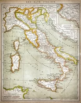
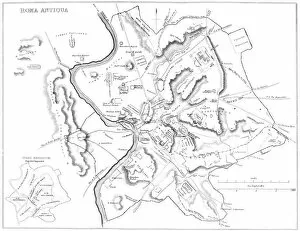
"Exploring the Past: Unveiling the Condition of Antique Maps" Step into a world where time stands still, as we delve into the captivating condition of antique maps. Each map tells a unique story, transporting us to distant lands and bygone eras. Gently unfurling an ancient scroll, we encounter an exquisite antique map of the Roman Empire. Its delicate lines depict a vast realm that once dominated Europe, offering a glimpse into its grandeur and complexity. Moving forward in time, our attention is drawn to another remarkable artifact - an antique map showcasing Britain under the Anglo Saxons. The intricate details reveal how this island nation evolved through centuries of cultural fusion and territorial shifts. A particularly intriguing piece catches our eye - a Falkland Islands Royal Engineer briefing map from 1982. This relic carries traces of history's conflicts etched upon its surface, reminding us of past struggles and their enduring impact on these remote islands. Venturing further across continents, we stumble upon an enchanting antique map depicting New Zealand in the 19th century. It unveils uncharted territories waiting to be explored while capturing the spirit of discovery that characterized this era. Not all maps have escaped unscathed from the ravages of time; one such example is a damaged antique map revealing Ireland's rich heritage amidst its visible wear and tear. Despite its imperfections, it serves as a testament to resilience and endurance throughout history. Journeying southward brings us face-to-face with an awe-inspiring antique map portraying Africa in 1873. Its faded hues evoke tales of exploration and colonization that shaped this vast continent for better or worse. Our gaze then falls upon yet another worn treasure - an aged depiction of Italy during the 19th century. Though marred by age-induced blemishes, it still manages to transport us back to a time when artistry flourished within Italian borders.