
Connective Tissue Collection (#4)
Connective tissue is a remarkable network that holds our bodies together, providing support and structure to various organs and systems


For sale as Licensed Images
Choose your image, Select your licence and Download the media
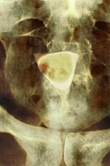
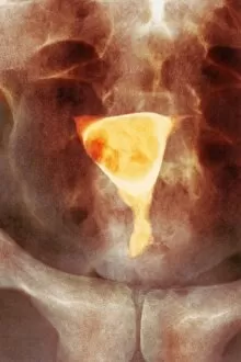
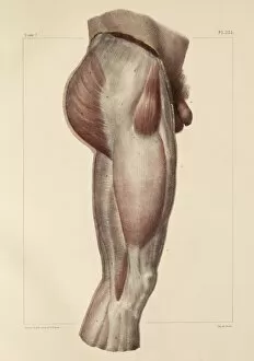
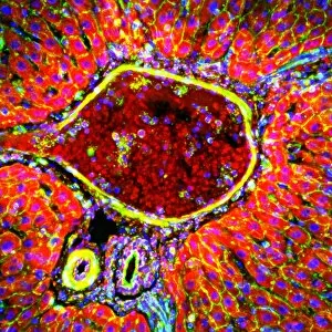
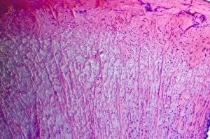
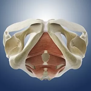
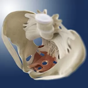
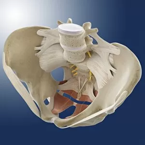
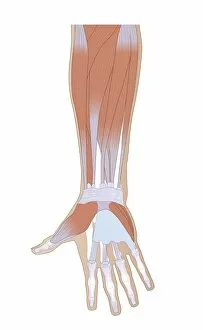
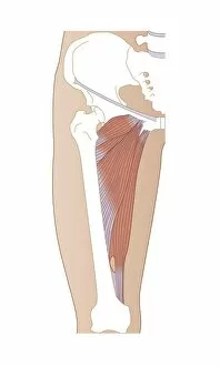
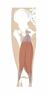
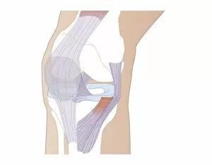
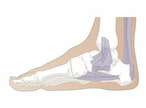
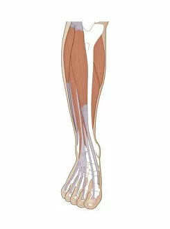
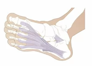
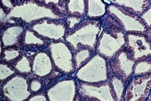
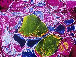
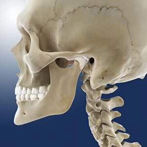
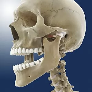
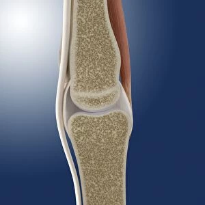
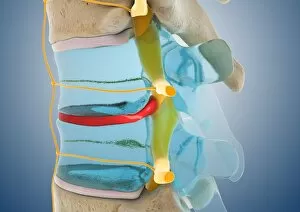
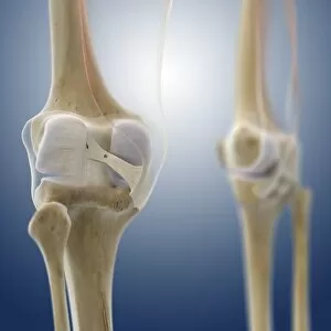
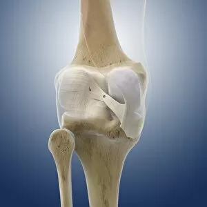
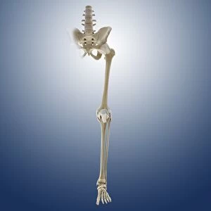
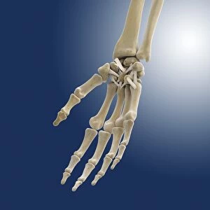
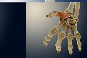
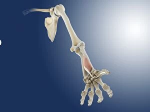
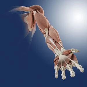
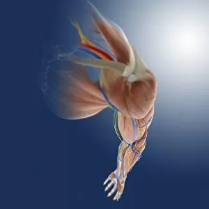
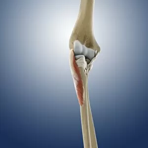
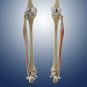
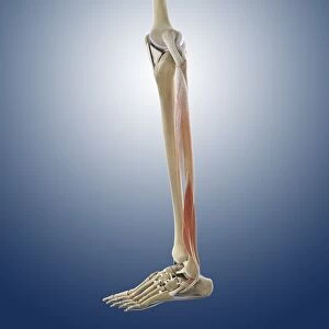
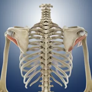
Connective tissue is a remarkable network that holds our bodies together, providing support and structure to various organs and systems. In the anatomy of the human knee joint, connective tissue plays a crucial role in maintaining stability and allowing smooth movement. Lactating breast tissue, as seen under a light microscope, showcases the intricate arrangement of connective fibers that aid in milk production. Examining tendons through scanning electron microscopy reveals their strong composition primarily made up of collagen fibers. These tough yet flexible strands provide resilience and enable efficient transmission of forces between muscles and bones. Artwork depicting outer ankle ligaments (C013 / 4452) illustrates how connective tissue safeguards joints from excessive movements while ensuring proper alignment during physical activities. Similarly, inner ankle ligaments (C013 / 4451) contribute to joint stability by connecting bones within the ankle region. Even on a microscopic level, connective tissue continues to amaze us. Computer artwork showcasing red blood cells highlights their vital role in transporting oxygen throughout our body via an intricate network of capillaries embedded within this specialized type of connective tissue. Delving into human tooth anatomy through artwork unveils another aspect where they are present – supporting structures like periodontal ligament that anchor teeth firmly within the jawbone. Fibroblast cells depicted in artwork demonstrate their pivotal function in synthesizing extracellular matrix components such as collagen and elastin – essential for wound healing and maintaining overall tissue integrity. Mesenchymal stem cells captured using scanning electron microscopy exhibit immense potential for regenerative medicine due to their ability to differentiate into various cell types found within different types of connective tissues. The optic nerve fibers imaged under SEM highlight how delicate yet resilient these structures are, responsible for transmitting visual information from our eyes to the brain with utmost precision thanks to surrounding supportive connective tissues. Lastly, examining fat tissue at high magnification reveals its unique structure composed mainly of adipocytes, which store energy and provide insulation.