
Coronaviridae Collection
"Unveiling the Intricate World of Coronaviridae: A Microscopic Glimpse into a Global Concern" In this captivating image


For sale as Licensed Images
Choose your image, Select your licence and Download the media
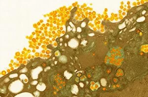
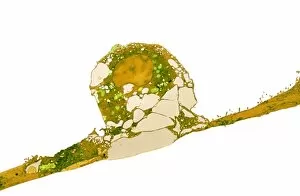
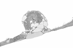
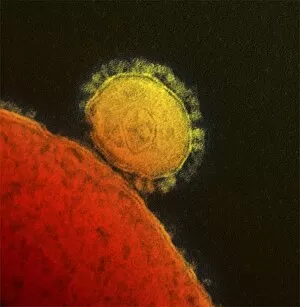
"Unveiling the Intricate World of Coronaviridae: A Microscopic Glimpse into a Global Concern" In this captivating image, we are granted a microscopic view of the notorious coronavirus, belonging to the family Coronaviridae. With its distinct crown-like appearance, it is no wonder why these viruses have garnered significant attention worldwide. This conceptual image beautifully illustrates the intricate structure and complexity of coronaviruses. Their spherical shape encapsulates genetic material that holds secrets to their infectious nature. As we delve deeper into their realm, we uncover various strains such as MERS coronavirus. Through transmission electron microscopy (TEM), scientists have captured stunning images like TEM C015 / 1772, TEM C015 / 1774, and TEM C015 / 1776 showcasing different angles and perspectives of MERS coronavirus. These high-resolution snapshots allow us to study their morphology in detail. The breathtaking TEM images continue with TEM C015 / 1773, TEM C015 / 7155, TEM C015 / 7157, and TEM C015 / 7158 providing further insight into the unique characteristics of MERS coronavirus. Each photograph unveils new information about its composition and behavior within host cells. As our understanding deepens through scientific exploration, researchers also present us with another remarkable capture - MERS coronavirus seen through TEM C017/8300. This particular image offers an even closer look at the virus's surface features and potential targets for therapeutic interventions. Coronaviridae has become more than just a scientific curiosity; it has emerged as a global concern due to recent outbreaks like COVID-19. The knowledge gained from studying these microscopic entities becomes crucial in combating future threats they may pose to human health. So let us marvel at these awe-inspiring visuals while appreciating the dedication of scientists who tirelessly work towards unraveling the mysteries surrounding coronaviridae – ultimately striving to protect and safeguard our world from their devastating impact.