
Cortical Collection
"Cortical: Unveiling the Intricacies of the Brain's Grey Matter" Delving into the depths of our complex neural system

For sale as Licensed Images
Choose your image, Select your licence and Download the media
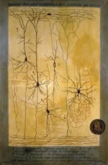
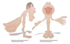
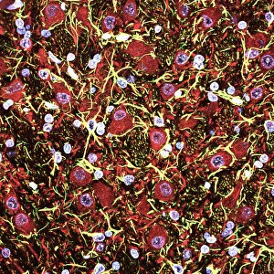
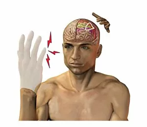
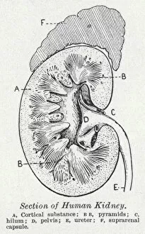
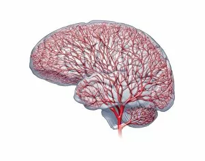
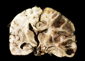
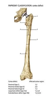
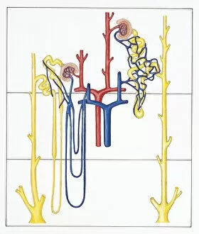
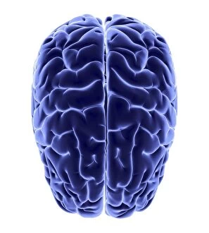
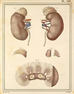
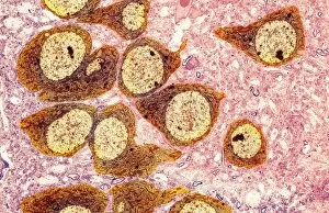
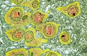
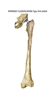
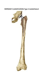
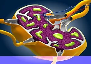
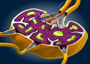
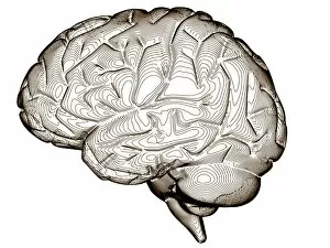
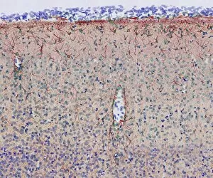
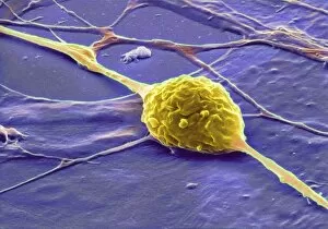
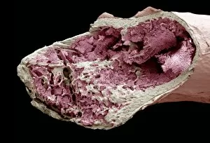
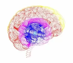
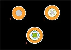
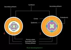
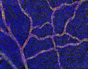
"Cortical: Unveiling the Intricacies of the Brain's Grey Matter" Delving into the depths of our complex neural system, cortical grey matter schema by Santiago Ramon Y Cajal reveals a mesmerizing tapestry of interconnected nerve cells. This intricate web forms the foundation of our cerebral cortex, orchestrating countless cognitive processes. Intriguingly, the motor homunculus model showcases how different body parts are represented in this cortical map. From tiny fingers to towering legs, each region corresponds to specific movements and sensations - an awe-inspiring testament to our brain's organization. Not limited to motor functions alone, sensory homunculi demonstrate how touch, taste, sight, and sound are meticulously processed within this remarkable structure, and is here that phantom pain after amputation arises; a haunting reminder that even when severed from reality, these cortical pathways continue to send signals. Beyond cognition lies another realm - one where litho depictions expose brain blood vessels as lifelines nourishing every corner of this enigmatic organ. Yet sometimes fate deals a cruel hand with brain injuries like C016 / 8920 or Paprosky femur defect classification C016 / 6621; reminders that even the most resilient cortex can be vulnerable. Meanwhile, illustrations depicting both cortical and juxtamedullary nephrons highlight their pivotal role in kidney function. These microscopic units filter waste and maintain homeostasis - an elegant dance orchestrated by nature itself since time immemorial. As we marvel at F006 / 6313 artwork portraying the intricacies of human brains or gaze upon ancient depictions showcasing kidney anatomy from 1825 – we are reminded once again of our perpetual quest for understanding. The mysteries held within these cortical realms beckon us closer towards unraveling their secrets while humbling us with their sheer complexity. Cortical: A gateway into humanity's exploration of its own mind and body – forever captivating and endlessly fascinating.