
Cranium Collection (page 3)
"Cranium: Unveiling the Mysteries of the Human Skull and Beyond" Delving into the depths of human anatomy


For sale as Licensed Images
Choose your image, Select your licence and Download the media
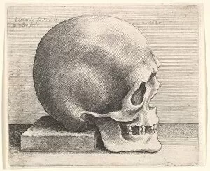
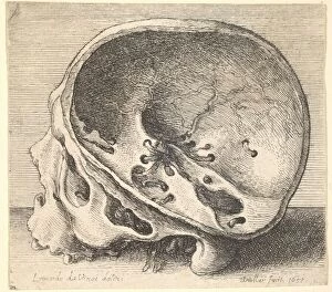
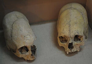
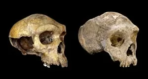
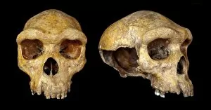
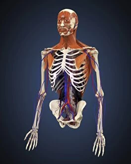
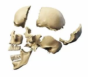
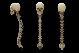
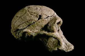
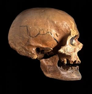
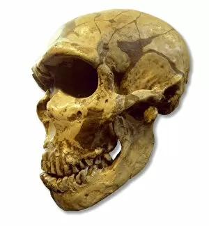
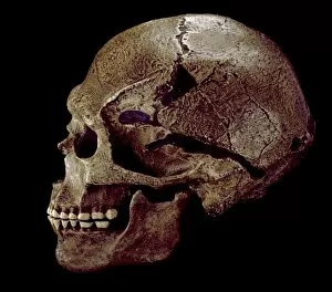
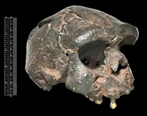
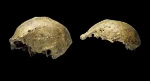
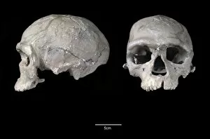
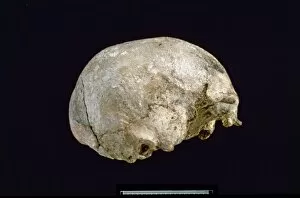
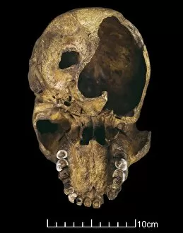
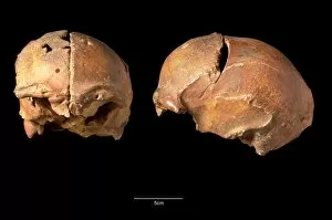
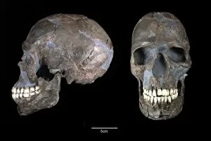
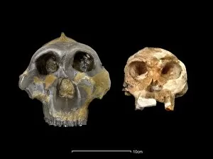
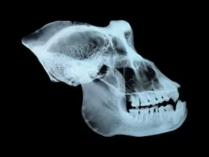
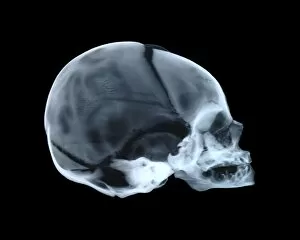
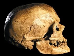
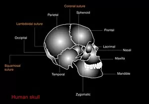
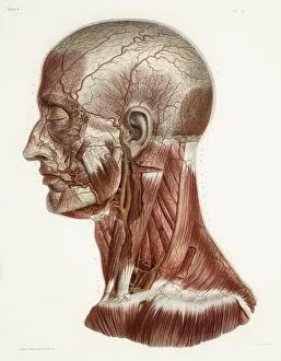
"Cranium: Unveiling the Mysteries of the Human Skull and Beyond" Delving into the depths of human anatomy, Leonardo da Vinci's intricate sketches of skull anatomy offer a glimpse into his fascination with unraveling the secrets held within our craniums. From his detailed drawings to modern-day advancements, we continue to explore this enigmatic structure. Hominid crania provide us with a window into our evolutionary past, showcasing how our ancestors' skulls have evolved over time. With full-body scans and MRI technology, scientists can now peer inside these ancient relics, uncovering clues about their lifestyles and adaptations. In a satirical twist on phrenology - an outdated pseudoscience that claimed personality traits could be determined by skull shape - we find humor in our quest for understanding. Yet amidst the satire lies an appreciation for how far we've come in deciphering the complexities of cranial morphology. The Paranthropus boisei (Zinjanthropus) cranium (OH5) takes us back millions of years, offering insights into early hominids' robust features. Meanwhile, X-ray images reveal intricate details of both human and animal skulls – from horses to primates – highlighting similarities and differences across species. One cannot ignore Sahelanthropus tchadensis; its fossilized skull provides a crucial link between apes and humans. This remarkable discovery challenges previous notions about our origins while fueling further exploration into humanity's beginnings. But it is not just scientific curiosity that draws attention to the cranium; it also holds significance in everyday life. Headaches become tangible through X-ray artwork, capturing both pain and beauty simultaneously. The human skull itself serves as a reminder of mortality but also showcases resilience throughout history. Finally, Homo erectus' Java Man cranium (Sangiran 17) cast allows us to step back in time once more – witnessing glimpses of ancient lives and pondering the mysteries that still surround our own existence.