
Cryptogamic Collection

Cryptogamic organisms are a fascinating world of microscopic wonders

Background imageCryptogamic Collection: Album ' Voyage en Ecosse Septembre 1880 ': Lake Katrine in Scotland

Album " Voyage en Ecosse Septembre 1880 ": Lake Katrine in Scotland
3562595 Album " Voyage en Ecosse Septembre 1880 ": Lake Katrine in Scotland; (add.info.: Scotland, Great Britain 29/09/1880 Photographer: Wilson, George Washington/g.w.w); Alinari Archives

Background imageCryptogamic Collection: Forest ferns

Forest ferns
3569141 Forest ferns; (add.info.: Great Britain 1867 Photographer: Allport); Alinari Archives, Florence

Background imageCryptogamic Collection: 'Debris': seaweed on the beach

"Debris": seaweed on the beach Italy. Date of Photograph:1950-1955

Background imageCryptogamic Collection: Forest vegetation at Loch Goil, Scotland

Forest vegetation at Loch Goil, Scotland Great Britain Scotland, Loch Goil. Date of Photograph:1867

Background imageCryptogamic Collection: Still life with fish, birds and vegetables

Still life with fish, birds and vegetables Great Britain. Date of Photograph:1865 ca

Background imageCryptogamic Collection: A forest plant

A forest plant Great Britain. Date of Photograph:1865 ca

Background imageCryptogamic Collection: Forest ferns

Forest ferns Great Britain. Date of Photograph:1867 ca

Background imageCryptogamic Collection: Pictorialist still life with mushrooms

Pictorialist still life with mushrooms. Date of Photograph:1934

Background imageCryptogamic Collection: Young girl photographed in a country road near the river Alcantara

Young girl photographed in a country road near the river Alcantara Taormina. Date of Photograph:26/04/1913

Background imageCryptogamic Collection: A stone bridge crosses a mountain stream, in Harz, Germany

A stone bridge crosses a mountain stream, in Harz, Germany. Germany Harz.. Date of Photograph:1890-1910 ca

Background imageCryptogamic Collection: Portrait of a woman sitting on a meadow next to mushrooms and asparagus in Vallombrosa

Portrait of a woman sitting on a meadow next to mushrooms and asparagus in Vallombrosa Reggello Vallombrosa. Date of Photograph:09/1922

Background imageCryptogamic Collection: Group of women gathering seaweed

Group of women gathering seaweed Japan. Date of Photograph:1885 ca

Background imageCryptogamic Collection: Algae emergeing from the water

Algae emergeing from the water. Date of Photograph:1965 ca

Background imageCryptogamic Collection: Close-up of Alsophila Australis cultivated in the garden of Prince Stigliano-Colonna in Naples

Close-up of Alsophila Australis cultivated in the garden of Prince Stigliano-Colonna in Naples Naples. Date of Photograph:1870 ca

Background imageCryptogamic Collection: 'Wild flowers and animals of Australia': mountain musk

"Wild flowers and animals of Australia": mountain musk Australia / Oceania. Date of Photograph:1910 ca

Background imageCryptogamic Collection: Full-length portrait of a girl, sitting on a low wall and surrounded by papier-mache doves

Full-length portrait of a girl, sitting on a low wall and surrounded by papier-mache doves. Her clothing and the setting of the portrait in the studio evoke a bucolic atmosphere

Background imageCryptogamic Collection: Microscopic enlargement of Achorion Schoenleinii, parasite fungus of man

Microscopic enlargement of Achorion Schoenleinii, parasite fungus of man. Date of Photograph:1897-1914 ca

Background imageCryptogamic Collection: Fabulous ringworm fungus on day 16 magnified under the microscope

Fabulous ringworm fungus on day 16 magnified under the microscope. Date of Photograph:1914 ca

Background imageCryptogamic Collection: Microscopic enlargment of Saccharomyces Equi

Microscopic enlargment of Saccharomyces Equi. Date of Photograph:1902-1913 ca

Background imageCryptogamic Collection: Saccharomyces apiculata: microscopic, unicellular fungus, of the Ascomycetes

Saccharomyces apiculata: microscopic, unicellular fungus, of the Ascomycetes, enlarged under a microscope. Date of Photograph:04/1913

Background imageCryptogamic Collection: Microscopic enlargment of Saccharomyces Cerevisiae

Microscopic enlargment of Saccharomyces Cerevisiae. Date of Photograph:04/1913

Background imageCryptogamic Collection: Phragmidium asperum fungus, belonging to the Uredineae, enlarged under a microscope

Phragmidium asperum fungus, belonging to the Uredineae, enlarged under a microscope. Date of Photograph:04/1914

Background imageCryptogamic Collection: Puccinia graminis: fungus belonging to the Uredineae, enlarged under a microscope

Puccinia graminis: fungus belonging to the Uredineae, enlarged under a microscope. Date of Photograph:1914

Background imageCryptogamic Collection: Fungus belonging to the Uredineae, called Uromyces Ficaria, enlarged under a microscope

Fungus belonging to the Uredineae, called Uromyces Ficaria, enlarged under a microscope. Date of Photograph:12/1902

Background imageCryptogamic Collection: Uredineae: parasite fungus that invades the Graminaceae producing the disease called rust

Uredineae: parasite fungus that invades the Graminaceae producing the disease called rust. Enlarged under a microscope. Date of Photograph:12/1902

Background imageCryptogamic Collection: Melampsora salicina fungus, belonging to the Uredineae class, enlarged under a microscope

Melampsora salicina fungus, belonging to the Uredineae class, enlarged under a microscope. Date of Photograph:12/1902

Background imageCryptogamic Collection: Puccinia arundinacea, belonging to the Hyphomycetes, enlarged under a microscope

Puccinia arundinacea, belonging to the Hyphomycetes, enlarged under a microscope. Date of Photograph:1898

Background imageCryptogamic Collection: Exidia auricularia fungus enlarged under a microscope

Exidia auricularia fungus enlarged under a microscope. Date of Photograph:12/1902

Background imageCryptogamic Collection: Capillitium of the Stemonitis ferruginea, fungus belonging to the Myxomycetes

Capillitium of the Stemonitis ferruginea, fungus belonging to the Myxomycetes. Enlarged under a microscope. Date of Photograph:12/1902

Background imageCryptogamic Collection: Phitosphora ingestans 'De Bary, ' the fungus responsible for the patato disease

Phitosphora ingestans "De Bary, " the fungus responsible for the patato disease. Enlarged under a microscope. Date of Photograph:12/1902

Background imageCryptogamic Collection: Capillitium and spores of the Bovista, a fungus belonging to the Gasteromycetes class

Capillitium and spores of the Bovista, a fungus belonging to the Gasteromycetes class. Enlarged under a microscope. Date of Photograph:12/1902

Background imageCryptogamic Collection: Peronospora viticola fungus enlarged under a microscope

Peronospora viticola fungus enlarged under a microscope. Date of Photograph:12/1902

Background imageCryptogamic Collection: Tuber cibarium fungus with spores, enlarged under a microscope

Tuber cibarium fungus with spores, enlarged under a microscope. Date of Photograph:1898

Background imageCryptogamic Collection: Penicillium glaucum: fungus belonging to the Perisporea species. Enlarged under a microscope

Penicillium glaucum: fungus belonging to the Perisporea species. Enlarged under a microscope. Date of Photograph:1898

Background imageCryptogamic Collection: Mucor Mucedo ascus (Mucorinea) enlarged under a microscope

Mucor Mucedo ascus (Mucorinea) enlarged under a microscope. Date of Photograph:1897

Background imageCryptogamic Collection: Alacodiscus formosus in the Diatomea, enlarged under a microscope

Alacodiscus formosus in the Diatomea, enlarged under a microscope. Date of Photograph:1914

Background imageCryptogamic Collection: Surirella gemma: yellow-brown alga belonging to the Diatomea

Surirella gemma: yellow-brown alga belonging to the Diatomea, and having free cells or cells in colonies with a silicified membrane, composed of two closed, box-like valves

Background imageCryptogamic Collection: Triceralium favus: yellow-brown alga belonging to the Diatomea

Triceralium favus: yellow-brown alga belonging to the Diatomea, and having free cells or cells in colonies with a silicified membrane, composed of two closed, box-like valves

Background imageCryptogamic Collection: Sphaeroplea annulina, belonging to the Sphaeropleacea family, enlarged under a microscope

Sphaeroplea annulina, belonging to the Sphaeropleacea family, enlarged under a microscope. Date of Photograph:1914

Background imageCryptogamic Collection: Ulothrix zonata, belonging to the Ulothrichea class, enlarged under a microscope

Ulothrix zonata, belonging to the Ulothrichea class, enlarged under a microscope. Date of Photograph:1914

Background imageCryptogamic Collection: Dictimopricom Grevilles (alga desmidiacea) enlarged under a microscope

Dictimopricom Grevilles (alga desmidiacea) enlarged under a microscope. Date of Photograph:1897

Background imageCryptogamic Collection: Uracnoide ornatus (Hyalodiscus) enlarged under a microscope

Uracnoide ornatus (Hyalodiscus) enlarged under a microscope. Date of Photograph:01/1890

Background imageCryptogamic Collection: Surirella gemma and Pleurosigma belonging to the Diatomea: yellow-brown algas with free cells or

Surirella gemma and Pleurosigma belonging to the Diatomea: yellow-brown algas with free cells or cells in colonies with a silicified membrane, composed of two closed, box-like valves

Background imageCryptogamic Collection: Microscopic enlargment of cells of Zygnema Cruciatum

Microscopic enlargment of cells of Zygnema Cruciatum. Date of Photograph:1914

Background imageCryptogamic Collection: Microscopic enlargement of vegetative cells of Cladophora Glomeratae (Rivularie)

Microscopic enlargement of vegetative cells of Cladophora Glomeratae (Rivularie). Date of Photograph:1914

Background imageCryptogamic Collection: Microscopic enlargment of cells of Oscillaria Viridis (Oscillatoria)

Microscopic enlargment of cells of Oscillaria Viridis (Oscillatoria). Date of Photograph:12/1902

Background imageCryptogamic Collection: Chara anteride (alga characea): male reproductive organ enlarged under a microscope

Chara anteride (alga characea): male reproductive organ enlarged under a microscope. Date of Photograph:1897

Background imageCryptogamic Collection: Common nostoc enlarged under a microscope

Common nostoc enlarged under a microscope. Date of Photograph:1914


For sale as Licensed Images

Choose your image, Select your licence and Download the media

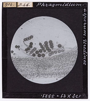
Cryptogamic organisms are a fascinating world of microscopic wonders. Take, for instance, the Achorion Schoenleinii fungus, a parasite that invades humans and causes ringworm. Under the microscope on day 16, this fabulous fungus reveals its intricate structure. But fungi come in all shapes and sizes. Saccharomyces Equi is a unicellular fungus belonging to the Ascomycetes family. When magnified under a microscope, it unveils its delicate beauty. Another member of the Saccharomyces family is Saccharomyces apiculata, an enchanting unicellular fungus that captivates with its microscopic details when observed closely. Moving on to other fungal marvels, Phragmidium asperum from the Uredineae class catches our attention. This rust-causing organism enlarges under the lens to reveal its complex structure. Uromyces Ficaria is another Uredineae species that invades Graminaceae plants and produces rust disease. Its enlarged form under a microscope showcases nature's intricacy at work. Melampsora salicina belongs to the same Uredineae class but offers unique characteristics when viewed up close. Its microscopic features tell tales of adaptation and survival in their own miniature universe. Puccinia arundinacea takes us into the realm of Hyphomycetes fungi with its enlarged appearance under a microscope. The complexity within this tiny organism leaves us in awe of nature's diversity. Lastly, we have Exidia auricularia—a captivating fungus whose enlargement under a microscope unravels its mysterious allure. Each detail invites us to explore further into this hidden world beneath our feet. Cryptogamic organisms offer glimpses into an unseen realm where beauty thrives at microscopic levels.