Crystalline Collection (#12)

"Captivating Crystalline Beauty

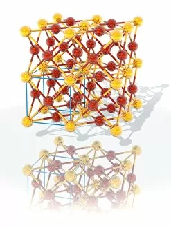
Background imageCrystalline Collection: Superconductor

Superconductor. Molecular model of the crystal structure of magnesium diboride (MgB2), a new super-conductor. Red spheres represent magnesium atoms and purple represent boron atoms

Background imageCrystalline Collection: Metal surface at the quantum level

Metal surface at the quantum level. This depiction of a metal surface is obtained by applying the Helium-3 spin-echo technique

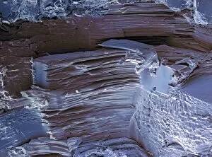
Background imageCrystalline Collection: Bismuth crystal

Bismuth crystal. Bismuth is a heavy, brittle, crystalline metal. This rectangular crystal structure only forms when bismuth is slowly cooled in the laboratory, never in nature

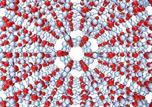
Background imageCrystalline Collection: Ice, molecular model

Ice, molecular model
Ice. Molecular model showing the hexagonal structure of ice. Atoms are represented as spheres and are colour-coded: hydrogen (white) and oxygen (red)

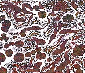
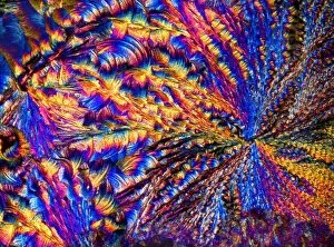
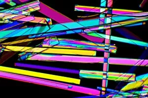
Background imageCrystalline Collection: Penicillin crystals, light micrograph

Penicillin crystals, light micrograph
Penicillin crystals. Polarised light micrograph of crystals of the antibiotic drug penicillin. Penicillin was the first antibiotic to be discovered

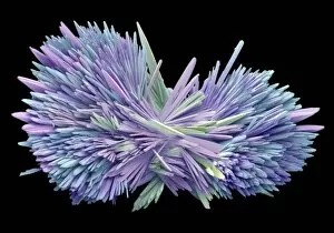
Background imageCrystalline Collection: Bronchodilator drug crystals, SEM

Bronchodilator drug crystals, SEM
Bronchodilator drug crystals, coloured scanning electron micrograph (SEM). These are crystals of the drug ventolin (salbutamol sulphate), a bronchodilator drug used to treat asthma

Background imageCrystalline Collection: Sea salt, SEM

Sea salt, SEM
Sea salt. Coloured scanning electron micrograph (SEM) of sea salt, showing its crystalline structure. Sea salt consists mainly of sodium chloride (NaCl), but unlike pure table salt

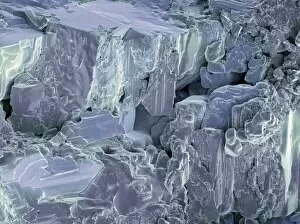
Background imageCrystalline Collection: Artificial sweetener crystals, SEM

Artificial sweetener crystals, SEM
Artificial sweetener crystals. Coloured scanning electron micrograph (SEM) of a freeze-fracture through an artificial sweetener tablet, showing its crystalline structure

Background imageCrystalline Collection: Sugar cube crystals, SEM

Sugar cube crystals, SEM
Sugar cube crystals. Coloured scanning electron micrograph (SEM) of a freeze-fracture through a sugar cube. The fracture plane shows the sucrose crystals that make up the sugar cube

Background imageCrystalline Collection: Silver nitrate crystals, LM

Silver nitrate crystals, LM
Silver nitrate crystals. Polarised light micrograph (LM) of silver nitrate (AgNO3) crystals. Silver nitrate is used in photography and as a test for chloride, bromide and iodide ions

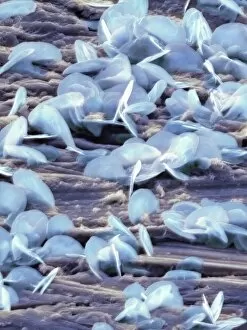
Background imageCrystalline Collection: Lead oxide crystals on lead, SEM

Lead oxide crystals on lead, SEM
Lead oxide crystals, coloured scanning electron micrograph (SEM). Lead is a heavy, soft, ductile metal that, under normal conditions, does not react with water

Background imageCrystalline Collection: Gold-copper alloy crystal

Gold-copper alloy crystal
Gold-copper alloy, molecular model. Gold atoms are gold, copper atoms are red. The crystal lattice of this alloy has a face centred cubic (FCC) structure

Background imageCrystalline Collection: Magnesium chloride crystals, LM

Magnesium chloride crystals, LM
Magnesium chloride. Polarised light micrograph (LM) of crystals of magnesium chloride (MgCl2). Typically, magnesium chloride forms complexes with other molecules; the hygroscopic hexahydrate

Background imageCrystalline Collection: Phenol crystals

Phenol crystals on a watchglass. Phenol (C6H5OH), an aromatic alcohol, is a crystalline solid. It dissolves in water, forming a weakly acid solution, carbolic acid. Phenol is corrosive and poisonous

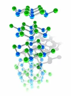
Background imageCrystalline Collection: Hexagonal boron nitride crystal

Hexagonal boron nitride crystal
Boron nitride crystal structure, molecular model. Boron atoms are green, nitrogen atoms are blue. Boron nitride is a compound that shares many properties with elemental carbon

Background imageCrystalline Collection: Tridymite crystal structure

Tridymite crystal structure, molecular model. Tridymite is a form of quartz (silicon dioxide) that forms at high temperatures

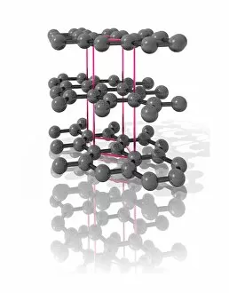
Background imageCrystalline Collection: Graphite

Graphite crystal. Computer model of the molecular structure of a graphite crystal. Graphite is used in pencil leads and as a lubricant

Background imageCrystalline Collection: Magnesium sulphate crystals, LM

Magnesium sulphate crystals, LM
Magnesium sulphate. Polarised light micrograph of crystals of magnesium sulphate (MgSO4), a chemical used medicinally and in the textile industry

Background imageCrystalline Collection: Zeolite crystals, light micrograph

Zeolite crystals, light micrograph
Zeolite crystals. Polarised light micrograph of crystals of zeolite from the Giants Causeway, Northern Ireland. Zeolites are hydrated aluminosilicate minerals and have a micro-porous structure

Background imageCrystalline Collection: Virus particles, conceptual artwork

Virus particles, conceptual artwork
Virus particles, conceptual computer artwork. Viruses contain a core of genetic material, either DNA (deoxyribonucleic acid) or RNA (ribonucleic acid) surrounded by a protein coat, or capsid

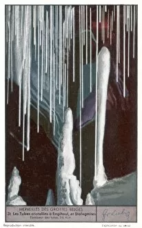
Background imageCrystalline Collection: Belgian Grotto 3

Belgian Grotto 3
Stalagmites and crystalline tubes at Engihoul


For sale as Licensed Images

Choose your image, Select your licence and Download the media

"Captivating Crystalline Beauty: A Glimpse into the Intricate World of Minerals and Molecules" Delicate copper and magnesium sulphate crystals under a powerful light microscope (LM) reveal their mesmerizing structure. Caffeine crystals, captured in a stunning light micrograph, showcase the intricate patterns formed by this stimulating compound. The perovskite crystal structure unveils its remarkable symmetry, creating an enchanting visual spectacle. A computer artwork depicts the brilliance of a diamond's crystalline lattice, reflecting its timeless allure. Cortisol crystals, magnified through a light micrograph (LM), exhibit their unique formation within our body's natural stress hormone. Vibrant copper sulphate crystals come to life when observed under a high-resolution light microscope (LM), revealing their captivating hues and shapes. The beach of Capo Coda Cavallo in Sardinia, Italy offers breathtaking views where nature's crystalline beauty meets pristine shores - truly an idyllic escape in Europe. Another glimpse into caffeine's microscopic world showcases its delicate crystal formations through an exquisite light micrograph. An artistic representation reveals the intricate patterns found within graphene sheets - highlighting both scientific marvels and aesthetic wonders. Baking soda crystals take on extraordinary shapes when examined using scanning electron microscopy (SEM), unveiling hidden textures that captivate the eye. Copper sulphate crystals shimmer with elegance as they are unveiled through the lens of a high-resolution light microscope (LM). A fly delicately perched on sugar granules is brought to life through scanning electron microscopy (SEM), showcasing how even everyday substances can possess unexpected beauty. In this collection of images and descriptions lies proof that there is unparalleled beauty hidden within every corner of our world – from minerals to molecules – reminding us to appreciate nature's artistry at both macroscopic and microscopic levels.