Cytoplasm Collection (#2)

"Cytoplasm: The Dynamic Hub of Cellular Activity" The cytoplasm, a bustling and vibrant region within the cell, plays a crucial role in various cellular processes

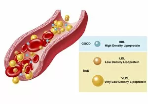
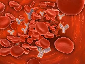
Background imageCytoplasm Collection: Good and bad cholesterol found in blood stream

Good and bad cholesterol found in blood stream

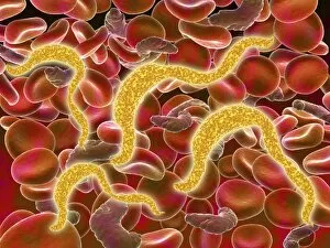
Background imageCytoplasm Collection: African trypanosomiasis in the red blood cells

African trypanosomiasis in the red blood cells. African trypanosomiasis, also known as sleeping sickness, is a parasitic infection caused by protozoa

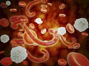
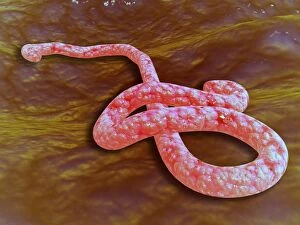
Background imageCytoplasm Collection: Conceptual image of ebola virus in blood stream

Conceptual image of ebola virus in blood stream

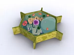
Background imageCytoplasm Collection: Conceptual image of a plant cell and its components

Conceptual image of a plant cell and its components

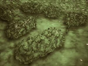
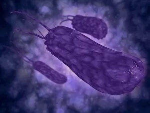
Background imageCytoplasm Collection: Microscopic view of E. coli bacteria cells

Microscopic view of E. coli bacteria cells
Microscopic view of Escherichia coli bacteria cells, commonly known as E. Coli. E. coli is a common type of bacteria that can get into food, such as beef and vegetables

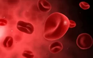
Background imageCytoplasm Collection: Microscopic view of red blood cells

Microscopic view of red blood cells

Background imageCytoplasm Collection: Acute coronary syndrome - microvascular obstruction

Acute coronary syndrome - microvascular obstruction

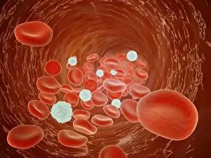
Background imageCytoplasm Collection: Red blood cells with leukocytes

Red blood cells with leukocytes

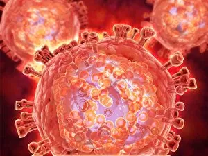
Background imageCytoplasm Collection: Cluster of HIV virus

Cluster of HIV virus. HIV is the human immunodeficiency virus that can lead to acquired immune deficiency syndrom, or AIDS

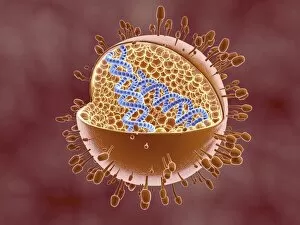
Background imageCytoplasm Collection: Cutaway view of Reoviridae virus showing dna inside

Cutaway view of Reoviridae virus showing dna inside. Reoviruses can affect the gastronintestinal system and respiratory tract

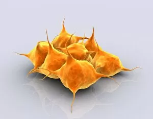
Background imageCytoplasm Collection: Conceptual image of a group of platelets

Conceptual image of a group of platelets

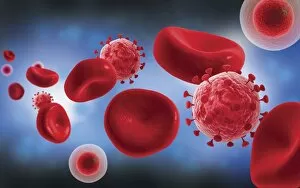
Background imageCytoplasm Collection: Microscopic view of blood cells with virus

Microscopic view of blood cells with virus

Background imageCytoplasm Collection: Microscopic view of Ebola virus

Microscopic view of Ebola virus (EBOV). EBOV causes extremely severe disease in humans and in non-human primates in the form of viral hemorrhagic fever

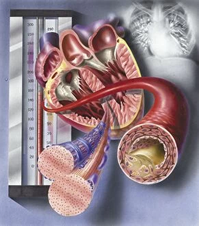
Background imageCytoplasm Collection: Interior view of heart with detail of muscle cells and atherosclerotic artery

Interior view of heart with detail of muscle cells and atherosclerotic artery. Chest x-ray in background
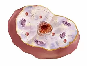
Background imageCytoplasm Collection: Human cell anatomy

Human cell anatomy

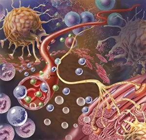
Background imageCytoplasm Collection: Nerve ending, seen in lower right, sends pain message from injured muscle

Nerve ending, seen in lower right, sends pain message from injured muscle. Blood vessel and immune cells are seen in the center and upper right of image

Background imageCytoplasm Collection: Thrombus forming on valve within vein

Thrombus forming on valve within vein

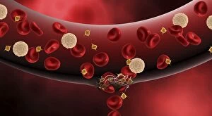
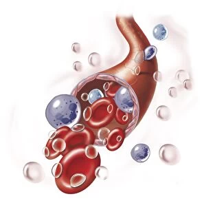
Background imageCytoplasm Collection: Blood vessel with platelets, white blood cells and red blood cells

Blood vessel with platelets, white blood cells and red blood cells

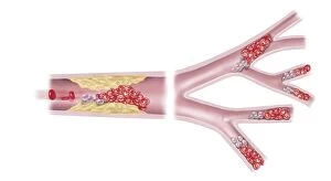
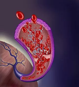
Background imageCytoplasm Collection: Normal artery compared to plaque and thrombus formation in artery

Normal artery compared to plaque and thrombus formation in artery

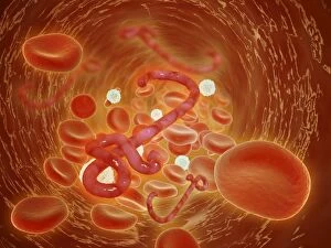
Background imageCytoplasm Collection: Conceptual image of ebola virus in artery

Conceptual image of ebola virus in artery

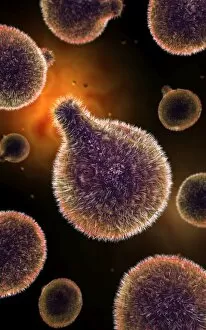
Background imageCytoplasm Collection: Microscopic view of poliovirus

Microscopic view of poliovirus. Poliovirus is the causative agent of polio, a human enterovirus. It is composed of an RNA genome and a protein capsid

Background imageCytoplasm Collection: Conceptual image of hemoglobin and red blood cells

Conceptual image of hemoglobin and red blood cells. Hemoglobin is a protein responsible for transporting oxygen in the red blood cells of vertebrates

Background imageCytoplasm Collection: Conceptual image of plasmodium causing malaria

Conceptual image of plasmodium causing malaria

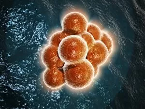
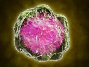
Background imageCytoplasm Collection: Microscopic view of a stem cell

Microscopic view of a stem cell. Stem cells are biological cells found in all multicellular organisms that can divide through mitosis and differentiate into diverse specialized cell types

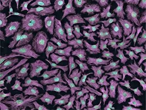
Background imageCytoplasm Collection: Multiphoton fluorescence image of HeLa cells

Multiphoton fluorescence image of HeLa cells with cytoskeletal microtubules (magenta) and DNA (cyan)

Background imageCytoplasm Collection: Red blood cell flow inside the artery

Red blood cell flow inside the artery

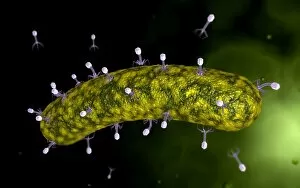
Background imageCytoplasm Collection: Microscopic view of bacteriophage attacking bacteria

Microscopic view of bacteriophage attacking bacteria

Background imageCytoplasm Collection: Conceptual image of Lactobacillus acidophilus

Conceptual image of Lactobacillus acidophilus. Lactobacillus is a gram-positive bacteria that occurs naturally in the human and animal gastrointestinal tract and mouth

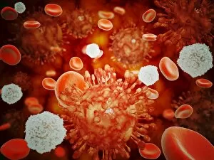
Background imageCytoplasm Collection: Microscopic view of H5N1 virus with red blood cells and white blood cells

Microscopic view of H5N1 virus with red blood cells and white blood cells

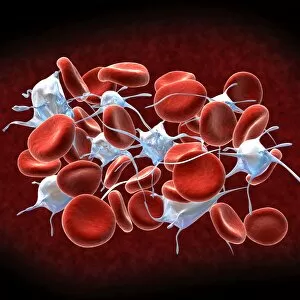
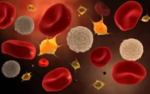
Background imageCytoplasm Collection: Conceptual image of platelets with red blood cells

Conceptual image of platelets with red blood cells
Conceptual image of platelets with white blood cells and red blood cells

Background imageCytoplasm Collection: Conceptual image of chromosomes inside the blood stream

Conceptual image of chromosomes inside the blood stream

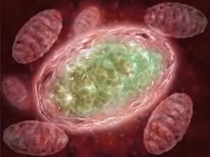
Background imageCytoplasm Collection: Microscopic view of Mitochondria

Microscopic view of Mitochondria. Mitochondria are the cells power producers. They convert energy into forms that are usable by the cell. Located in the cytoplasmcells activities

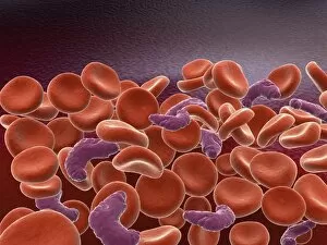
Background imageCytoplasm Collection: Conceptual image of sickle cell anemia with red blood cells

Conceptual image of sickle cell anemia with red blood cells

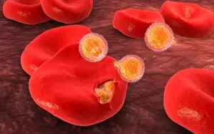
Background imageCytoplasm Collection: Conceptual image of malaria parasites within red blood cells

Conceptual image of malaria parasites within red blood cells

Background imageCytoplasm Collection: Conceptual image of a red blood cell

Conceptual image of a red blood cell

Background imageCytoplasm Collection: Conceptual image of a platelet

Conceptual image of a platelet

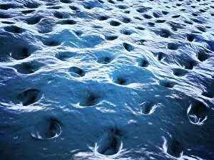
Background imageCytoplasm Collection: Microscopic view of Dentine

Microscopic view of Dentine. Dentine is the main supporting structure of the tooth and is the second hardest tissue in the body after enamel. It is 70% mineral and acellular

Background imageCytoplasm Collection: Normal artery versus artery in spasm

Normal artery versus artery in spasm

Background imageCytoplasm Collection: Conceptual image of plasmodium

Conceptual image of plasmodium

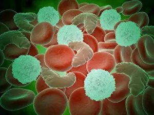
Background imageCytoplasm Collection: Sickle cell anemia with red blood cells and white bood cells

Sickle cell anemia with red blood cells and white bood cells
Conceptual image of sickle cell anemia with red blood cells and white bood cells

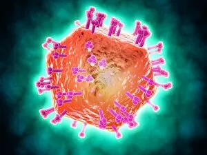
Background imageCytoplasm Collection: Conceptual image of HIV virus

Conceptual image of HIV virus. HIV is the human immunodeficiency virus that can lead to acquired immune deficiency syndrome, or AIDS

Background imageCytoplasm Collection: Conceptual image of cytoskeleton

Conceptual image of cytoskeleton. The cytoskeleton is a cellular scaffolding or skeleton contained within a cells cytoplasm, and is present in all cells

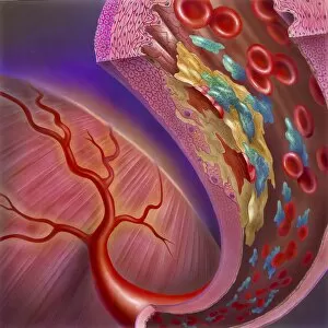
Background imageCytoplasm Collection: Artery showing atherosclerotic plaque, platelets and red blood cells

Artery showing atherosclerotic plaque, platelets and red blood cells
Close-up of artery showing atherosclerotic plaque, platelets and red blood cells

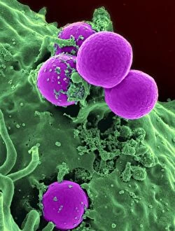
Background imageCytoplasm Collection: Scanning electron micrograph of a human neutrophil ingesting MRSA

Scanning electron micrograph of a human neutrophil ingesting MRSA

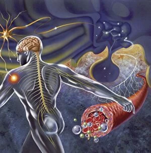
Background imageCytoplasm Collection: Schematic of the hypothalamus receiving nerve impulses from the body

Schematic of the hypothalamus receiving nerve impulses from the body and sending messages to the circulatory and nervous system

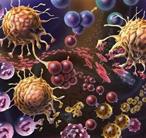
Background imageCytoplasm Collection: Artistic representation of the immune systems reaction to bacteria invading the tissues

Artistic representation of the immune systems reaction to bacteria invading the tissues

Background imageCytoplasm Collection: Arteriole with red blood cells, white blood cells and oxygen

Arteriole with red blood cells, white blood cells and oxygen

Background imageCytoplasm Collection: Conceptual image of helicobacter

Conceptual image of helicobacter. Helicobacter is a genus of gram-negative bacteria possessing a characteristic helix shape


For sale as Licensed Images

Choose your image, Select your licence and Download the media

"Cytoplasm: The Dynamic Hub of Cellular Activity" The cytoplasm, a bustling and vibrant region within the cell, plays a crucial role in various cellular processes. Through advanced techniques like Transmission Electron Microscopy (TEM) and fluorescent micrograph imaging, scientists have unraveled its intricate functions. In nerve cells, myelination of nerve fibers is a remarkable phenomenon that enhances signal transmission. TEM images reveal the mesmerizing beauty of these intricately wrapped fibers, highlighting the importance in maintaining efficient communication within our nervous system. During cell division, another captivating process captured through fluorescent micrographs, the cytoplasm orchestrates an elaborate dance to ensure accurate distribution of genetic material. These stunning visuals showcase how this dynamic organelle actively participates in life's fundamental cycle. Exploring further into TEM imagery unveils plasma cells with their distinctive appearance. Cytoplasmic structures are clearly visible as they produce antibodies vital for immune defense against pathogens such as human respiratory syncytial virus – depicted here under microscopic view. Fascinatingly, not only does cytoplasm support specialized cells but also aids viruses themselves. In AIDS infections visualized by TEM, we witness viral particles budding from host cells' cytoplasms – shedding light on the intricate relationship between pathogen and host. Delving deeper into cellular architecture reveals an illustration showcasing the nucleus containing essential components like mitochondria units, DNA strands coiled into chromosomes – all enveloped within a sea of cytoplasm. This conceptual image emphasizes its pivotal role in preserving genetic information and supporting vital cellular functions. Finally, a captivating artwork depicts platelets alongside red and white blood cells – symbolizing harmony within our circulatory system. The presence of these diverse cell types underscores how each relies on well-coordinated interactions facilitated by the ever-active cytoplasm. Whether it be myelination in nerve fibers or aiding viral replication or participating in cell division, the cytoplasm emerges as a dynamic and indispensable hub of cellular activity.