
Cytosol Collection
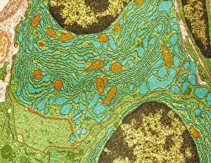
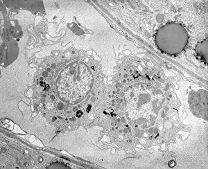
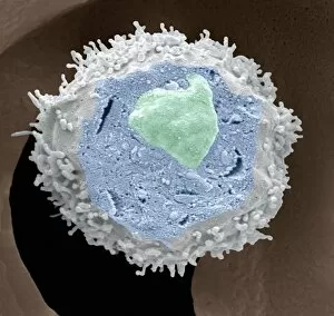
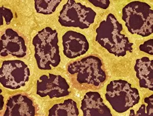
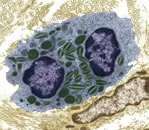
Cytosol: The Dynamic Fluid of Cellular Life Plasma cells, TEM images showcasing their intricate structure and functionality

For sale as Licensed Images
Choose your image, Select your licence and Download the media
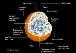
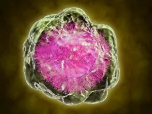
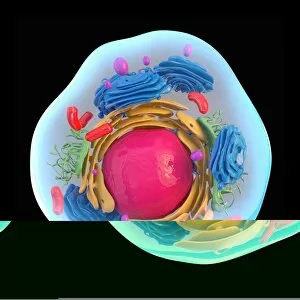
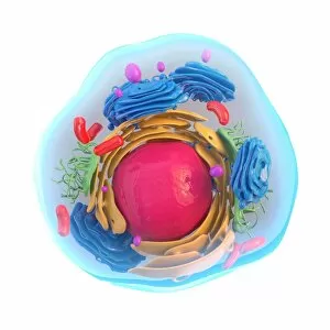
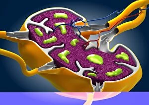
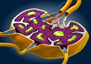
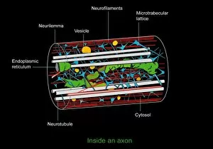
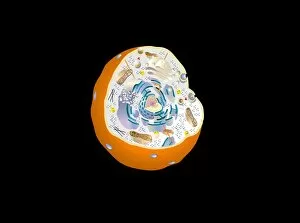
Cytosol: The Dynamic Fluid of Cellular Life Plasma cells, TEM images showcasing their intricate structure and functionality, are just one example of the wonders that occur within the cytosol. This dynamic fluid, found in all living animal cells including lymphocytes and macrophage white blood cells captured beautifully through SEM and TEM techniques, is a bustling hub of activity. Imagine a conceptual image of the cytoskeleton - an intricate network resembling a city's infrastructure. It provides structural support to maintain cell shape while also facilitating cellular movement and transportation. Within this framework lies the cytosol - a watery solution containing various molecules such as proteins, ions, sugars, and enzymes. Macrophage white blood cells are key players in our immune system's defense mechanism. Through TEM imagery capturing their detailed morphology from different angles, we can appreciate how they navigate through tissues like sentinels on high alert for any foreign invaders or damaged cells. In artwork depicting animal cells (such as C013/9985), we get a glimpse into the complexity hidden beneath their seemingly simple exterior. The cytosol acts as a medium where vital metabolic reactions take place; it serves as both an energy source and storage facility for essential molecules required for cellular processes. Lymph nodes (artwork C013/4632) play a crucial role in filtering harmful substances from our body fluids. Embedded within these nodes are lymphocyte white blood cells (SEM image), which patrol tirelessly through the cytosol-filled channels seeking out potential threats to neutralize them effectively. The concept may seem abstract at first glance but understanding its significance brings us closer to unraveling life's mysteries at the cellular level. From plasma cells to macrophages, from lymph nodes to animal cell artwork – each piece contributes to our knowledge about this vibrant fluid that sustains life itself.