
Dermal Collection
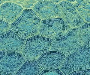
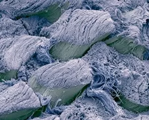
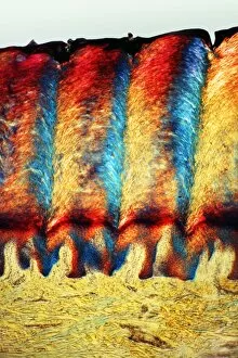
"Dermal Wonders: Exploring the Intricacies of Skin and Its Marvels" 1️⃣ Zebra fish skin under the SEM


For sale as Licensed Images
Choose your image, Select your licence and Download the media
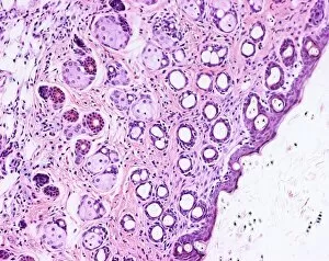
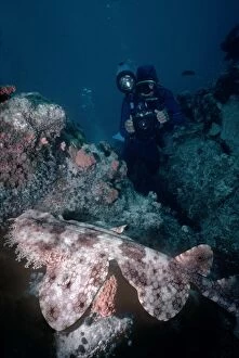
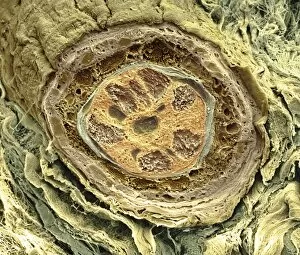
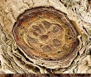
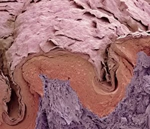
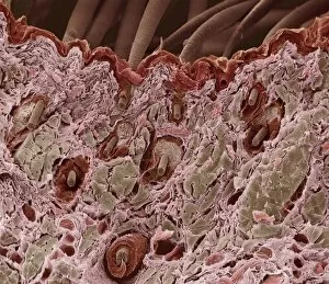
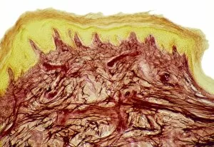
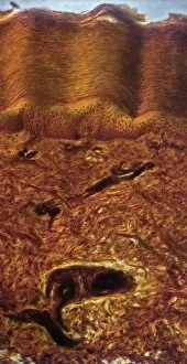
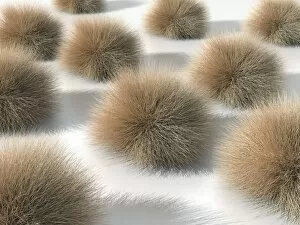
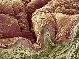
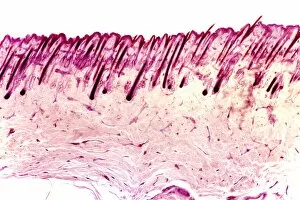
"Dermal Wonders: Exploring the Intricacies of Skin and Its Marvels" 1️⃣ Zebra fish skin under the SEM: Unveiling the intricate patterns and textures of this aquatic creature's dermal layer. 2️⃣ Psoriasis: A closer look at the enigmatic skin condition, revealing its impact on the dermis and beyond. 3️⃣ Pigmented skin mole C013 / 7318: Delving into the fascinating world of moles, their unique pigmentation, and their connection to our dermal landscape. 4️⃣ Skin and hair follicles in a mesmerizing light micrograph: Witnessing nature's artistry as we explore the interplay between our epidermis and hair growth. 5️⃣ Picture No. 10767831 captures Echinus esculentus, an edible sea urchin with stunning radial rows bumps adorning its upper surface. Nature's beauty knows no bounds. 6️⃣ Cutaneous leishmaniasis in 1917: Reflecting on historical records to understand how this parasitic infection affects our precious dermal layers. 7️⃣ Hair follicle magnified under SEM C014 / 0382 & C014 / 0383: Peering into these microscopic wonders that play a vital role in our hair growth cycle. 8️⃣ Hairy scalp skin illuminated through a light micrograph: Appreciating the complexity of our scalp's structure as it supports lush locks atop our heads. 9️⃣ Skin structure depicted through artwork C016 / 7541: Merging science with creativity to portray the intricacies of human dermis like never before – a masterpiece within ourselves. 🔟 Finger skin revealed under SEM lens: Discovering unseen details etched onto every inch of our fingertips – where touch meets sensation.