
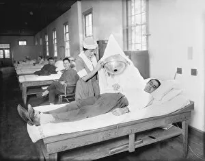
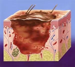
Dermatological Collection (#2)
"Dermatological Wonders: Exploring the Artistry of Skin Disorders" Delve into the intricate world of dermatology, where skin disorders become captivating works of art


For sale as Licensed Images
Choose your image, Select your licence and Download the media
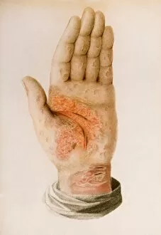
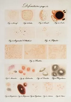
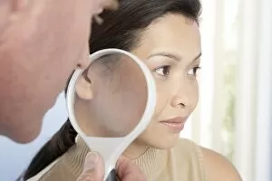
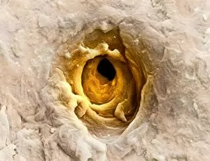
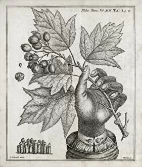
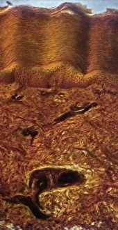
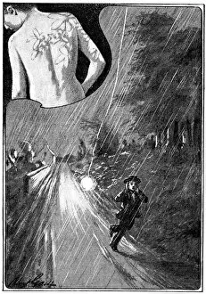
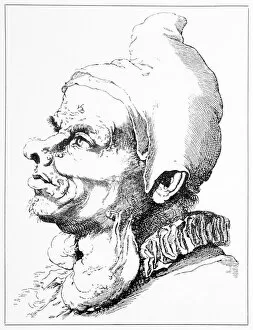
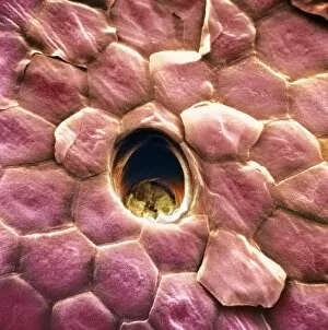
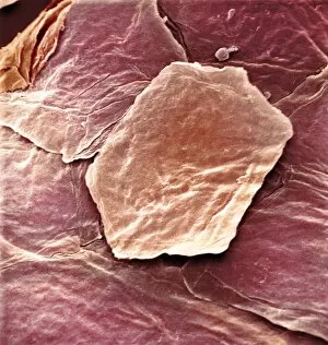
"Dermatological Wonders: Exploring the Artistry of Skin Disorders" Delve into the intricate world of dermatology, where skin disorders become captivating works of art. From illustrations showcasing human skin to coloured engravings depicting various conditions, this collection unveils the beauty and complexity that lies beneath our surface. Step into a realm where science meets aesthetics as we examine the mesmerizing images captured by a dermatoscope. These close-up views reveal the intricacies of our skin's topography, resembling an otherworldly landscape under scanning electron microscopy (SEM). Travel back in time through historical engravings from renowned medical books. Witness Psoriasis brought to life in vibrant colours by Robert Willan in 1808 and Favus meticulously depicted by Daniel Cornelius Danielssen in 1892. These illustrations serve as timeless reminders of how far dermatology has come. Discover advertisements from yesteryears promoting hypo-allergenic soaps like Viola Cream, designed to soothe and nourish troubled skin. The vivid lithographs transport us to an era when skincare was both a necessity and an indulgence. Uncover rare conditions such as Congenital Ichthyosis, immortalized in G. M. Olbers' 1830 engraving. Marvel at Baron Jean Louis Alibert's depiction of Ringworm from his 1838 book – a testament to early attempts at understanding and treating these perplexing ailments. Join us on this visual journey through centuries past, where art intertwines with medicine to shed light on the mysteries residing within our largest organ – the skin. Let these captivating images ignite your curiosity about dermatology's rich history while appreciating its profound impact on human health and well-being.