
Dermatology Collection
"Dermatology: Exploring the Canvas of Skin Disorders and Artwork" Discovering the intricate world unveils a captivating blend of science, artistry, and innovation


For sale as Licensed Images
Choose your image, Select your licence and Download the media
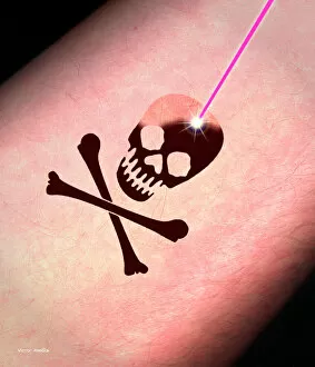
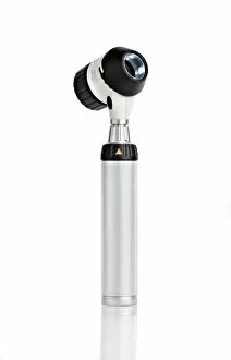
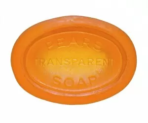
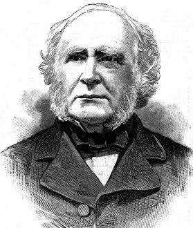
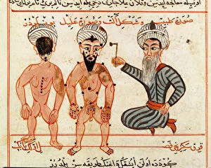
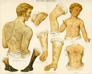
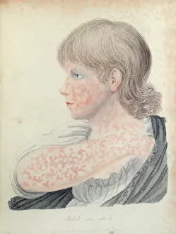
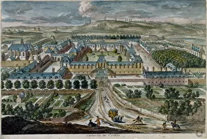
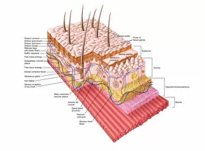
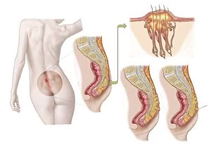
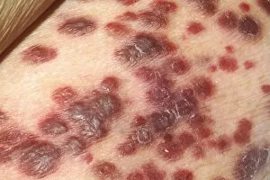
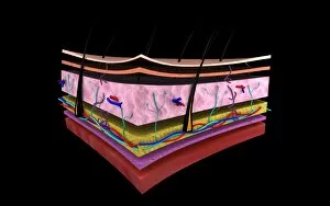
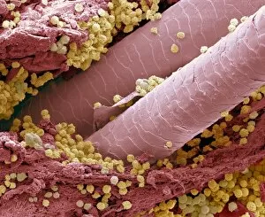
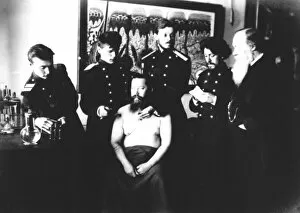
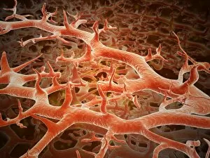
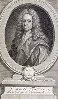
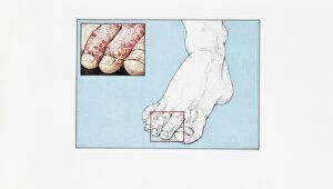
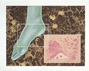
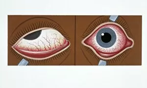
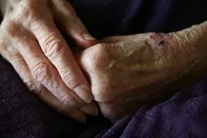
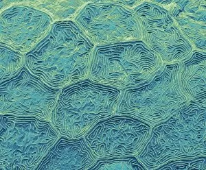
"Dermatology: Exploring the Canvas of Skin Disorders and Artwork" Discovering the intricate world unveils a captivating blend of science, artistry, and innovation. From ancient practices to modern advancements, this field delves into the realm of skin disorders while showcasing stunning artwork that celebrates its beauty. In our quest for healthy skin, hypo-allergenic soap emerges as a gentle companion. With its soothing touch, it caters to those with sensitive skin, offering respite from irritations and allergies. The evolution also witnesses remarkable breakthroughs like laser tattoo removal. As ink fades away under precise beams of light, individuals can embark on a fresh canvas for self-expression. Peering through the lens of a dermatoscope reveals an extraordinary view - an up-close exploration of the skin's surface in astonishing detail. This tool allows professionals to diagnose various conditions accurately while unraveling mysteries hidden beneath our epidermis. Zooming further into nature's wonders brings us Zebra fish skin captured by scanning electron microscopy (SEM). Its mesmerizing patterns remind us that even within tiny scales lies breathtaking complexity waiting to be discovered. Tracing back centuries ago, we stumble upon Ms Sup Turc 693 fol. 46v - an exquisite vellum depicting cauterization treatment for leprosy lesions in 1466. This historical artifact showcases early attempts at combating severe skin disorders with meticulous care and precision. Fast forward to Saint-Louis Hospital in the early 17th century; an engraving transports us back in time where dedicated surgeons and dermatologists worked tirelessly to heal their patients' ailments. Their commitment echoes through generations as they paved the way for modern-day advancements. A glimpse into history wouldn't be complete without Sir Erasmus Wilson gracing us with his presence through a Punch cartoon engraving. A renowned English surgeon and dermatologist, he left an indelible mark on this field with his expertise and contributions.