
Diagnostics Collection

"Diagnostics: Unveiling the Inner Workings of Our Health" In the realm of medical diagnostics

Background imageDiagnostics Collection: Medieval urine wheel

Medieval urine wheel. Labelled in Latin, this 15th century diagram shows some of the possible colours of urine (outer edge of circle) to help doctors diagnose urine-related diseases

Background imageDiagnostics Collection: Colour Blindness Test

Colour Blindness Test
Colour blindness test. Computer artwork of an Ishihara colour test card used to check for red- green colour blindness. The card is comprised of red and orange dots on a background of green dots

Background imageDiagnostics Collection: Full body scan, MRI scan

Full body scan, MRI scan
Full body scan of a female swimmer. Her brain is purple (top), her heart and arteries are red/orange (upper centre), her kidneys are pale green (centre)

Background imageDiagnostics Collection: Brain anatomy, MRI scan

Brain anatomy, MRI scan
Brain anatomy. Coloured magnetic resonance imaging (MRI) scan of the human head from the side. The sagittal scan has halved the brain, revealing the internal anatomy

Background imageDiagnostics Collection: Eye examination

Eye examination
MODEL RELEASED. MODEL RELEASED. Eye examination. Teenage (16 year old) girl looks through a refractor during an eye examination at an ophthalmologist

Background imageDiagnostics Collection: Normal EEG read out of the brains alpha waves

Normal EEG read out of the brains alpha waves
Normal electroencephalogram (EEG) read-out of the brains alpha waves. original is b/w m400/01m

Background imageDiagnostics Collection: Ruptured breast implants, MRI

Ruptured breast implants, MRI
Ruptured breast implants. Coloured magnetic resonance imaging (MRI) scan of ruptures in both of a womans breast implants (red). These are intracapsular ruptures, the most common type

Background imageDiagnostics Collection: Phrenology bust

Phrenology bust. Phrenology was a popular pseudoscience of the nineteenth century. The basis for the theory lay in a misplaced belief that a persons character

Background imageDiagnostics Collection: Neck, side view, coloured X-ray

Neck, side view, coloured X-ray
Neck from the side, coloured X-ray. The front of the body is at right. The spine runs down centre. It is made up of block-like bones called vertebrae

Background imageDiagnostics Collection: Slipped disc

Slipped disc. Spinal model demonstrating a slipped intervertebral disc. The slipped disc is the small bulge (red, centre), near the base of the spine

Background imageDiagnostics Collection: Those who tremble at the hand of a doctor, illustration from

Those who tremble at the hand of a doctor, illustration from
STC445985 Those who tremble at the hand of a doctor, illustration from The Works of Hippocrates, 1934 (colour litho) by Kuhn-Regnier

Background imageDiagnostics Collection: Dental plaque and tartar

Dental plaque and tartar. Clinical view of a the teeth in a patients lower jaw, with the incisors coated in bacterial plaque and tartar (brown)

Background imageDiagnostics Collection: Broken arm bone, digital X-ray

Broken arm bone, digital X-ray
Broken arm bone. Digital frontal X-ray of the fractured right humerus (upper arm bone) of a patient in their early twenties

Background imageDiagnostics Collection: Normal hand, digital X-ray

Normal hand, digital X-ray
Normal hand. Digital X-ray of the normal right hand of a patient in their late twenties. Various sets of data relating to the image can be seen around the edges of the X-ray

Background imageDiagnostics Collection: Radiologist examining knee MRI scans C014 / 1300

Radiologist examining knee MRI scans C014 / 1300
MODEL RELEASED. Radiologist examining knee MRI (magnetic resonance imaging) scans

Background imageDiagnostics Collection: Sheep skull

Sheep skull. Coloured X-ray of a sagittal section through a sheeps (Ovis aries) skull. Sheep have 32 permanent teeth; 12 molars and eight incisors on the bottom jaw and 12 molars on the top jaw

Background imageDiagnostics Collection: Cardiac stress test, artwork

Cardiac stress test, artwork
Cardiac stress test. Artwork showing a patient undergoing a cardiac stress test on a treadmill. At top are ECG (electrocardiogram) traces showing the patients heart rate at rest (left)

Background imageDiagnostics Collection: Brain scan, conceptual image C017 / 7392

Brain scan, conceptual image C017 / 7392
MODEL RELEASED. Brain scan, conceptual composite image

Background imageDiagnostics Collection: Brain scan, conceptual image C017 / 7394

Brain scan, conceptual image C017 / 7394
MODEL RELEASED. Brain scan, conceptual composite image

Background imageDiagnostics Collection: Brain scan, conceptual image C017 / 7398

Brain scan, conceptual image C017 / 7398
MODEL RELEASED. Brain scan, conceptual composite image

Background imageDiagnostics Collection: Brain scan, conceptual image C017 / 7397

Brain scan, conceptual image C017 / 7397
MODEL RELEASED. Brain scan, conceptual composite image

Background imageDiagnostics Collection: Brain scan, conceptual image C017 / 7391

Brain scan, conceptual image C017 / 7391
MODEL RELEASED. Brain scan, conceptual composite image

Background imageDiagnostics Collection: Light microscope F006 / 7165

Light microscope F006 / 7165
Light microscope on a laboratory bench

Background imageDiagnostics Collection: Light microscope F006 / 7167

Light microscope F006 / 7167
Light microscope on a laboratory bench

Background imageDiagnostics Collection: Biological research F006 / 7189

Biological research F006 / 7189
Biological research

Background imageDiagnostics Collection: Light microscope F006 / 7186

Light microscope F006 / 7186
Light microscope on a laboratory bench

Background imageDiagnostics Collection: Light microscope F006 / 7190

Light microscope F006 / 7190
Light microscope

Background imageDiagnostics Collection: Light microscope F006 / 7166

Light microscope F006 / 7166
Light microscope on a laboratory bench

Background imageDiagnostics Collection: ECG, artwork F006 / 4606

ECG, artwork F006 / 4606
ECG. Computer artwork of an electrocardiogram (ECG) showing a normal heart rate

Background imageDiagnostics Collection: Light microscope eyepieces F006 / 7188

Light microscope eyepieces F006 / 7188
Light microscope eyepieces

Background imageDiagnostics Collection: Light microscope F006 / 7184

Light microscope F006 / 7184
Light microscope on a laboratory bench

Background imageDiagnostics Collection: Blood samples F007 / 8178

Blood samples F007 / 8178
Blood samples

Background imageDiagnostics Collection: Urine samples F007 / 8175

Urine samples F007 / 8175
Urine samples

Background imageDiagnostics Collection: Urine and blood samples F007 / 8179

Urine and blood samples F007 / 8179
Urine and blood samples

Background imageDiagnostics Collection: Heart monitoring, conceptual image F008 / 3202

Heart monitoring, conceptual image F008 / 3202
Heart monitoring, conceptual image

Background imageDiagnostics Collection: Food science laboratory F008 / 3276

Food science laboratory F008 / 3276
MODEL RELEASED. Food science laboratory

Background imageDiagnostics Collection: Light microscope F008 / 2043

Light microscope F008 / 2043
Light microscope

Background imageDiagnostics Collection: Medical equipment and drugs F008 / 3189

Medical equipment and drugs F008 / 3189
Medical equipment and drugs

Background imageDiagnostics Collection: Microscopy F008 / 2042

Microscopy F008 / 2042
Microscopy

Background imageDiagnostics Collection: Blood smear F008 / 2176

Blood smear F008 / 2176
MODEL RELEASED. Blood smear

Background imageDiagnostics Collection: Medical equipment and drugs F008 / 3190

Medical equipment and drugs F008 / 3190
Medical equipment and drugs

Background imageDiagnostics Collection: Blood pressure pills, conceptual image F008 / 3174

Blood pressure pills, conceptual image F008 / 3174
Blood pressure pills, conceptual image

Background imageDiagnostics Collection: Biological research F008 / 2174

Biological research F008 / 2174
MODEL RELEASED. Biological research

Background imageDiagnostics Collection: Biological research F008 / 2175

Biological research F008 / 2175
MODEL RELEASED. Biological research

Background imageDiagnostics Collection: Woman using handheld machine F008 / 2179

Woman using handheld machine F008 / 2179
MODEL RELEASED. Woman using handheld machine

Background imageDiagnostics Collection: Biological research F008 / 2178

Biological research F008 / 2178
MODEL RELEASED. Biological research

Background imageDiagnostics Collection: Medical equipment and drugs F008 / 3188

Medical equipment and drugs F008 / 3188
Medical equipment and drugs

Background imageDiagnostics Collection: Heart monitoring, conceptual image F008 / 3204

Heart monitoring, conceptual image F008 / 3204
Heart monitoring, conceptual image


For sale as Licensed Images

Choose your image, Select your licence and Download the media

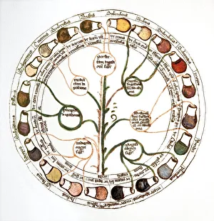
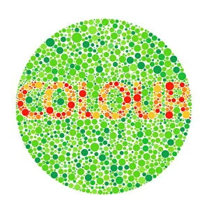
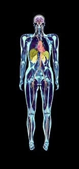
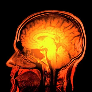
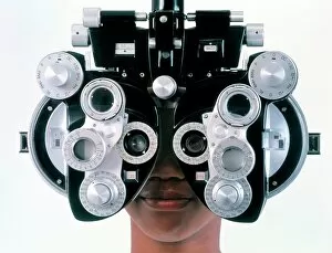
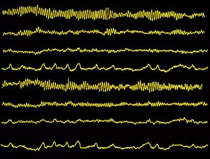
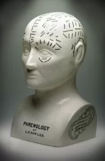
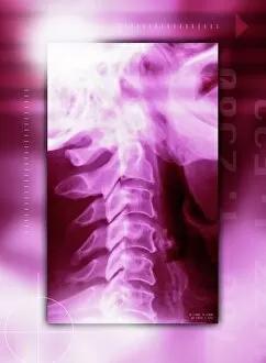
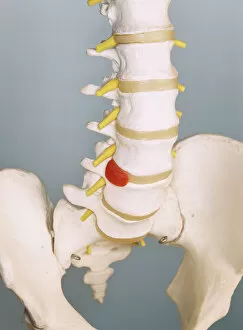
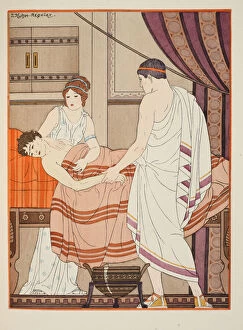
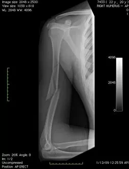
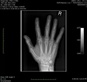
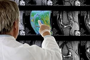
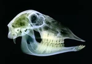
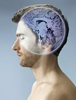
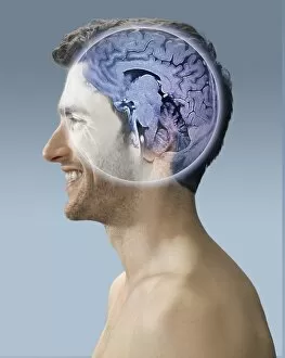
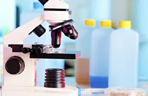
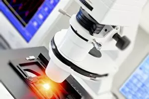
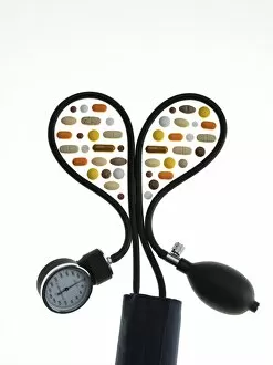
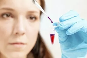
"Diagnostics: Unveiling the Inner Workings of Our Health" In the realm of medical diagnostics, a journey through time and technology reveals our quest to decipher the mysteries hidden within our bodies. From medieval urine wheels to cutting-edge scans, each diagnostic tool has played a crucial role in unraveling ailments and guiding us towards better health. Color blindness test charts have allowed us to understand how individuals perceive colors differently, shedding light on this fascinating condition that affects many. Meanwhile, full-body scans and MRI scans have revolutionized medical imaging by providing detailed glimpses into our innermost structures – from brain anatomy to ruptured breast implants. The enigmatic workings of the human mind are unraveled through EEG readings showcasing normal alpha waves, offering insights into cognitive processes. Eye examinations not only help determine visual acuity but also aid in detecting underlying conditions that may impact vision. A phrenology bust reminds us of an era when studying bumps on one's head was believed to reveal personality traits – a captivating yet debunked practice. However, modern neck X-rays provide valuable information about slipped discs and other spinal abnormalities with their vivid colored images. As we delve deeper into diagnostics, electrocardiograms (ECGs) become essential tools for assessing heart health by measuring its electrical activity. The intricate beauty of the human heart is unveiled as these tests guide physicians towards accurate diagnoses and effective treatments. From ancient practices rooted in curiosity to state-of-the-art technologies pushing boundaries, diagnostics continue to evolve alongside medical advancements. Each technique serves as a window into understanding our bodies' complexities while empowering healthcare professionals with invaluable knowledge necessary for healing and well-being.