
Diaphragm Collection (#5)
The diaphragm, a remarkable muscle located beneath the lungs, plays a vital role in our body's functioning


For sale as Licensed Images
Choose your image, Select your licence and Download the media
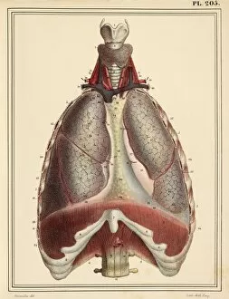
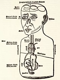
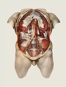
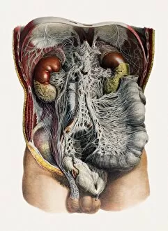
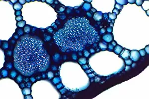
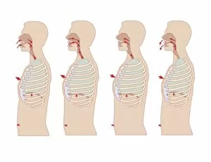
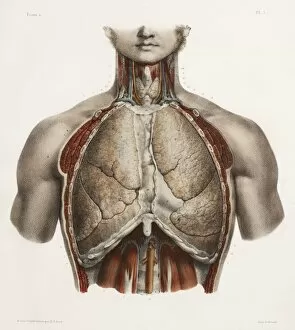
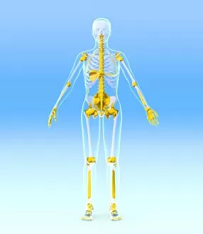
The diaphragm, a remarkable muscle located beneath the lungs, plays a vital role in our body's functioning. From chakras and the nervous system to the cardiovascular system, this powerful muscle impacts various aspects of our well-being. In historical artwork, we can witness depictions of the diaphragm as part of intricate anatomical drawings. These illustrations not only showcase its position but also highlight its significance in respiration mechanics. A diagram reveals how it moves down during exhalation and up during inhalation, aiding us in breathing effortlessly. One notable example is Victor Stiebel's dress design inspired by the diaphragm. This unique creation beautifully represents both fashion and anatomy, merging artistry with science seamlessly. Moving beyond respiration, artwork portraying the human digestive system sheds light on how the diaphragm interacts with other bodily functions. It demonstrates that this muscle influences digestion by maintaining proper pressure within our abdominal cavity. Delving deeper into its complexity, microscopic images reveal nerve synapses within the diaphragm through transmission electron microscopy (TEM). These intricate connections emphasize its crucial role in transmitting signals throughout our body. Even as early as 1892, advertisements for Dollond's new diaphragm shutter showcased innovative uses for this muscular marvel outside of biology. This invention revolutionized photography by controlling exposure time accurately. Exploring further into human anatomy unveils an elaborate network connecting the respiratory system and internal organs specifically designed for males. Such detailed diagrams provide invaluable insights into how these structures work harmoniously to sustain life. Taking a broader perspective view of the human body reveals an awe-inspiring sight: whole organs and bones coexisting alongside complex systems like nerves, lymphatic vessels, and blood vessels. This comprehensive illustration showcases just how intricately interconnected everything truly is within us. A midsection view allows us to explore internal organs more closely—providing insight into their placement relative to one another while highlighting their individual roles.