
Diatoms Collection (page 2)
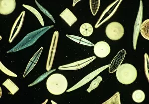
Diatoms, the mesmerizing art of nature's microscopic algae, captivate our imagination with their intricate beauty


For sale as Licensed Images
Choose your image, Select your licence and Download the media
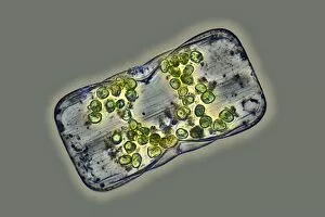
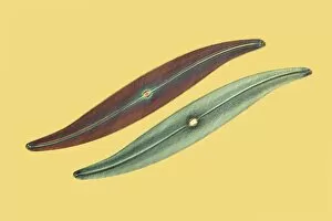
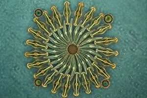
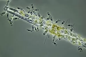
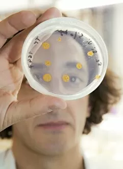
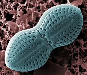
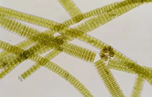
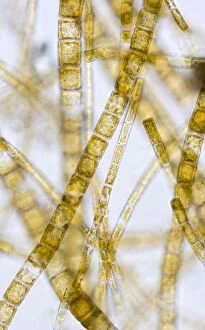
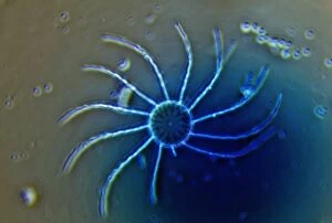
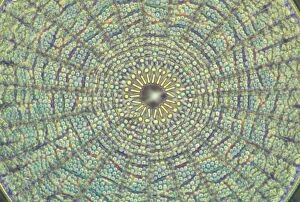
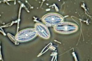
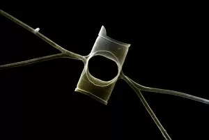
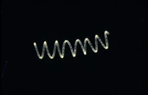
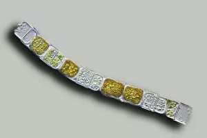
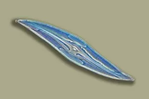
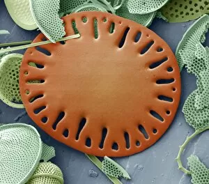
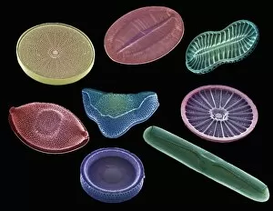
Diatoms, the mesmerizing art of nature's microscopic algae, captivate our imagination with their intricate beauty. Ernst Haeckel, a renowned biologist and artist, immortalized these diatom wonders through his stunning illustrations. From marine plankton samples to fossilized remains, they have left an indelible mark on our understanding of the natural world. Under the lens of a microscope at 25 times magnification, diatoms reveal themselves as delicate structures floating in the vastness of the ocean. Their symmetrical patterns and vibrant hues create a kaleidoscope that enchants scientists and artists alike. In Picture No. 11675478, we witness a close-up view captured by scanning electron microscopy (SEM). This technique allows us to explore their intricate details with unparalleled precision. The result is an awe-inspiring image that showcases the complexity hidden within these seemingly simple organisms. Fossilized diatoms provide us with glimpses into ancient ecosystems long gone. SEM imaging reveals their preserved forms, reminding us of their role as key players in Earth's history. These tiny creatures have witnessed countless changes over millions of years and continue to shape our planet today. The interplay between diatoms and blue-green algae is another captivating sight revealed by SEM imagery. In this microcosmic world, different species coexist harmoniously, creating breathtaking scenes reminiscent of abstract art. As we delve deeper into this microscopic realm through SEM technology, we uncover more secrets held by these remarkable organisms. Each new image brings forth fresh perspectives on their structure and function—reminding us that even in minuscule forms lies immense beauty waiting to be discovered. From Ernst Haeckel's artistic renderings to cutting-edge scientific techniques like SEM imaging—diatoms never cease to amaze us with their elegance and importance in shaping our planet's past and present. Through these captivating visuals, we are reminded once again that beauty can be found even in the tiniest corners of our natural world.